Have you ever stopped to consider just how complex your fingers actually are? The anatomy of finger structures is a marvel of biological engineering, combining bones, ligaments, tendons, muscles, nerves, and blood vessels into an incredibly precise system that allows you to perform tasks ranging from threading a needle to lifting heavy objects. Understanding hand joints anatomy can help you appreciate normal function, recognize when something feels wrong, and communicate more clearly with healthcare providers.
Whether you’re an artist learning to draw hands accurately, a student studying for anatomy exams, or simply someone who wants to understand their own body better, this guide covers the key structures. You’ll explore knuckle anatomy, learn the differences between the anatomy of a finger at each segment, and discover how the anatomy of the finger works as an integrated system.
Bones of the Finger
The Three Phalanges Each finger (except the thumb) contains three bones called phalanges: the proximal phalanx (closest to the hand), the middle phalanx, and the distal phalanx (the fingertip bone). The thumb has only two phalanges — proximal and distal — which is why it moves differently and offers such strong pinching force.
The bones of the anatomy of finger structures are relatively slender and become progressively smaller toward the fingertip. Fractures anywhere along the phalanges are common injuries; understanding which bone is affected helps determine the appropriate treatment and recovery protocol.
The Metacarpals and Their Role
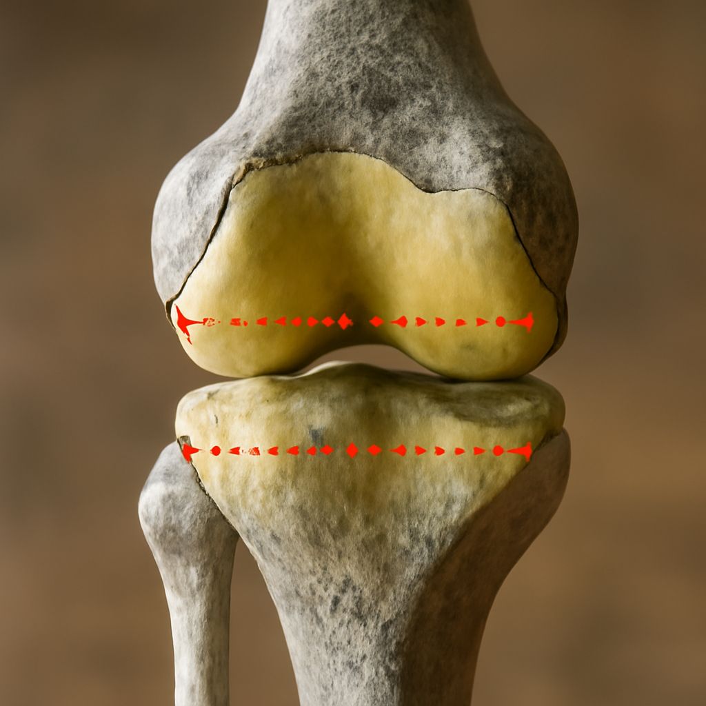
Though not technically finger bones, the five metacarpals form the palm of the hand and connect to the proximal phalanges at the metacarpophalangeal (MCP) joints. These MCP joints are what you commonly think of as the knuckles — the prominent bumps you see when you make a fist. Detailed hand joints anatomy begins right here at the MCP level, where the finger meets the hand proper.
Knuckle Anatomy: The Joints of the Finger
MCP Joints
Knuckle anatomy at the MCP level involves a condyloid joint that allows flexion, extension, abduction, and adduction. This flexibility gives you the ability to spread your fingers wide and bring them together, as well as to bend and straighten them. Strong collateral ligaments on either side of the joint prevent excessive sideways movement.
PIP and DIP Joints
The proximal interphalangeal (PIP) joint sits at the middle knuckle of each finger and is a hinge joint — it moves primarily in one plane, allowing flexion and extension. The distal interphalangeal (DIP) joint at the outermost knuckle operates similarly. Understanding knuckle anatomy at both the PIP and DIP levels is essential for diagnosing finger injuries, since each joint is vulnerable to different types of sprains, dislocations, and degenerative changes.
Tendons, Ligaments, and Soft Tissue
Flexor and Extensor Tendons
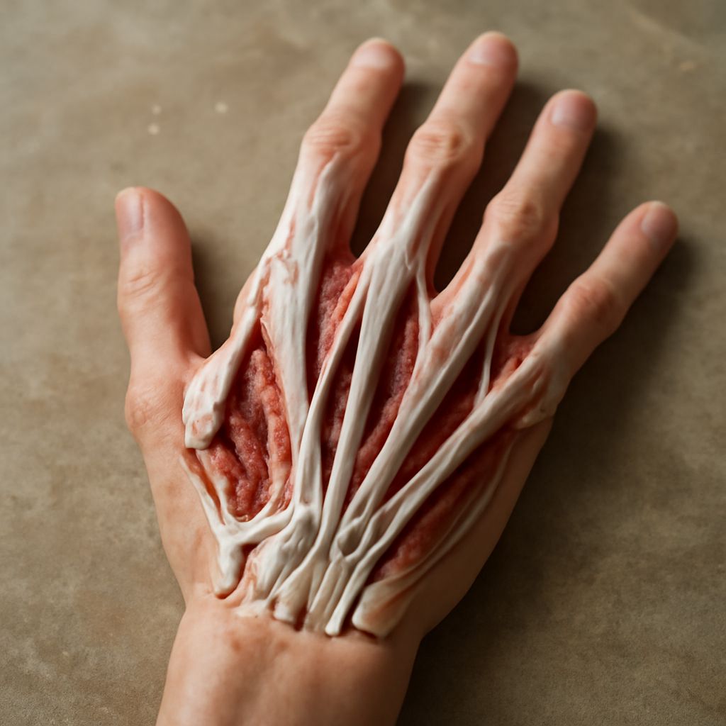
The anatomy of a finger would be incomplete without discussing its tendons. Flexor tendons run along the palm side of the finger and pull the digits into a curled position. Extensor tendons run along the back of the hand and straighten the fingers. These tendons travel through narrow fibrous sheaths that keep them close to the bone, allowing efficient force transmission from the forearm muscles that control finger movement.
Ligaments and Pulleys
A series of annular and cruciate pulleys hold the flexor tendons in position against the bone. Damage to these pulleys — common in rock climbers — causes the tendon to bowstring away from the bone, significantly reducing grip strength. The anatomy of the finger’s ligamentous system is a key focus area for hand surgeons and physical therapists treating finger injuries.
Nerves and Blood Supply
Sensation and motor control in the fingers come primarily from three nerves: the median nerve, the ulnar nerve, and the radial nerve. The median nerve supplies the thumb, index finger, middle finger, and half of the ring finger; the ulnar nerve covers the other half of the ring finger and the little finger. Understanding the nerve distribution across the anatomy of the finger helps clinicians pinpoint where a nerve injury or compression has occurred.
Blood supply arrives through the digital arteries running along each side of the finger. These vessels are small but critical; even minor lacerations can cause significant bleeding, and damage to digital arteries can compromise the health of the fingertip tissue downstream.
Bottom line: The anatomy of finger structures — from bones and joints to tendons and nerves — is a beautifully coordinated system that enables remarkable dexterity. Reviewing hand joints anatomy and knuckle anatomy gives you a meaningful foundation for understanding common injuries and why proper hand care matters. If you experience persistent pain or reduced range of motion in any finger joint, consult a qualified healthcare professional for evaluation.