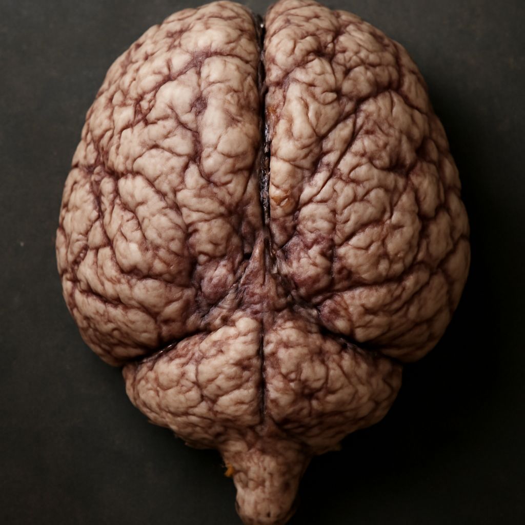
Have you ever wondered what makes a sheep’s brain unique, or how its structure compares to your own? The study of sheep brains offers a fascinating look into animal anatomy, revealing not just the intricate workings of these animals, but also broader principles shared among mammals. By examining a sheep brain diagram and diving into the layout of sheep brains in animal anatomy, you can better understand the functional architecture that underlies behavior and adaptation in the animal kingdom.
Whether you’re a student, educator, or simply curious about biological systems, exploring sheep brains in animal anatomy can provide a hands-on way to grasp core anatomical concepts. The sheep brain diagram is more than just a visual aid—it’s a roadmap to understanding how brains are organized, how different regions interact, and how the study of sheep brains can illuminate the inner workings of mammalian nervous systems.
Introduction to Sheep Brains and Their Importance
Sheep brains are frequently used in anatomical studies due to their manageable size, relative similarity to human brains, and availability. In animal anatomy, they serve as a valuable model for teaching about neural structures and basic brain functions. The study of sheep brains allows you to visualize the three-dimensional organization of the mammalian brain, bridging the gap between textbook diagrams and real-world biological forms.
This accessibility makes sheep brains ideal subjects for dissection and exploration. Their structure includes all the major regions found in more complex brains, yet in a form that fits comfortably in your hand. Through this lens, sheep brains in animal anatomy enable you to see first-hand the organization of essential regions like the cerebrum, cerebellum, and brainstem.
Learning to interpret a sheep brain diagram equips you with foundational skills for understanding neuroanatomy across species. You’ll discover not only the visible differences between sheep and human brains, but also the surprising similarities that point to shared evolutionary origins. Recognizing these features in sheep brains can demystify the complexity of nervous systems in general.
Exploring the Sheep Brain Diagram: Key Structures
An accurate sheep brain diagram is your primary tool for navigating this organ’s landscape. It typically displays the major external and internal features, including the two hemispheres of the cerebrum, the cerebellum located at the back, and the stalk-like brainstem connecting to the spinal cord. By examining these features in the sheep brain diagram, you gain an appreciation for how form and function are intertwined in animal anatomy.
Within the sheep brain diagram, you’ll find labels for critical structures like the corpus callosum, optic chiasm, thalamus, hypothalamus, and olfactory bulbs. Each of these parts plays a specific role in processing sensory input, coordinating movement, and regulating internal functions. As you study these anatomical elements, you not only learn their locations but also their relationships to one another.
Understanding the sheep brain diagram also involves recognizing the spatial orientation of these structures. The longitudinal fissure, for example, divides the two hemispheres, while the cerebellum’s distinctive ridges and the elongated brainstem become easily identifiable landmarks. By repeatedly referencing the sheep brain diagram during investigation or dissection, you solidify your ability to identify these features both visually and conceptually.
As you move from diagrams to hands-on work, partial features and alternative views—such as coronal and sagittal sections—enhance your understanding further. These perspectives allow you to connect textbook knowledge with practical anatomy, making the study of sheep brains in animal anatomy a truly immersive learning experience.
Comparing Sheep Brains with Human and Other Mammalian Brains
One of the most valuable learning opportunities when examining sheep brains in animal anatomy is drawing comparisons with other mammals, particularly humans. The overall layout found on a sheep brain diagram closely mirrors that of the human brain, reflecting shared evolutionary pathways and core neural functions. Both brains possess a cerebrum, cerebellum, brainstem, and similar internal structures, although the proportions and surface complexity differ.
The primary distinction between sheep and human brains lies in the development and folding (or gyrification) of the cerebrum. In humans, the cerebrum is much larger relative to body size and features more convolutions, which allow for increased surface area and cognitive complexity. On a sheep brain diagram, you’ll notice smoother cerebral hemispheres—a reflection of a less complex but still remarkably functional brain.
Despite these differences, examining sheep brains provides insights into the fundamental organization that supports sensory processing, motor control, and basic cognitive tasks across species. The shared features, such as the placement of the thalamus and hypothalamus, highlight conserved mechanisms that have stood the test of evolutionary time. By appreciating both the similarities and differences, you gain a deeper understanding of how structure supports function in mammalian neural systems.
Comparisons with other animal species—such as rodents, primates, or even larger livestock—can further enrich your grasp of animal anatomy. Each species exhibits adaptations that reflect its environment and behavioral needs, and the sheep brain diagram becomes a reference point for identifying these evolutionary trends.
Functions of Major Regions in the Sheep Brain Diagram
To fully appreciate the sheep brain diagram, it is important to connect the labeled structures to their functional roles. The cerebrum, the largest part shown on the diagram, is responsible for integrating sensory input and initiating voluntary movement. In sheep, this region processes information related to vision, hearing, touch, and even the complex olfactory cues that guide their behavior.
The cerebellum, situated at the rear of the brain, plays a crucial part in coordinating balance and smooth, coordinated movement. Its unique surface, often depicted in the sheep brain diagram as ridged or foliated, reflects its ability to handle intricate motor tasks—essential for grazing animals that must navigate uneven terrain.
The brainstem, including the midbrain, pons, and medulla oblongata, ensures survival by controlling vital involuntary processes such as heart rate, breathing, and digestion. In sheep brains in animal anatomy, these foundational systems remain highly conserved, underscoring their importance for life-sustaining functions across all mammals.
Other regions like the thalamus and hypothalamus direct sensory information to appropriate processing centers and maintain internal balance. Tracing these pathways using a sheep brain diagram helps reinforce your anatomical knowledge and illustrates the elegance of neural networks in action.
Educational Uses and Dissection of Sheep Brains
The study of sheep brains in animal anatomy is a cornerstone of biology and neuroscience education. Dissection activities, guided by a detailed sheep brain diagram, allow you to translate abstract anatomical concepts into tangible, real-world observations. By carefully examining the brain’s exterior and slicing through key planes, you identify firsthand the structures described in texts and lectures.
Hands-on dissection fosters a deeper understanding of spatial relationships and the complexity of neural tissue. Using proper technique and referencing the sheep brain diagram throughout the process ensures accuracy and enhances learning. You’ll learn to distinguish regions by texture, color, and connectivity, building a sensory memory of the brain’s organization.
Many educational settings also incorporate partial dissections or virtual models, supplementing physical exploration with digital tools. These interactive resources help reinforce concepts and accommodate various learning styles. Whether working on your own or as part of a class, the experience of investigating sheep brains in animal anatomy is both informative and memorable.
Ethical sourcing and proper specimen handling are integral parts of these activities. Practicing respect for the animal and maintaining cleanliness during dissection underscores the responsibility inherent in studying biological systems.
Conclusion: Why Study the Sheep Brain Diagram?
Engaging with the sheep brain diagram and exploring sheep brains in animal anatomy equips you with a solid foundation in neuroanatomy. This experience links theoretical understanding with practical skills, deepening your appreciation for both the complexity and commonality of mammalian nervous systems. By comparing, identifying, and analyzing these structures, you open the door to broader studies in biology, veterinary science, and comparative anatomy.
Next steps: Continue building your anatomical expertise by reviewing diagrams, participating in guided dissections, and exploring virtual brain models. Practice identifying key regions repeatedly, and always prioritize safe, ethical handling of specimens. Each session brings you closer to mastering the intricacies of animal brains and prepares you for more advanced explorations in the fascinating world of neuroanatomy.