Horse Cartoons, Horse Skeleton Anatomy, and Horse Anatomy Quiz Guide
What happens when you combine a love of horses with a need to understand them more deeply? You end up studying everything from playful horse cartoons that capture equine personality to detailed horse skeleton anatomy charts that explain why a horse moves the way it does. Whether you are an illustrator looking to draw horses more convincingly, a student preparing for an horse anatomy quiz, or an equestrian who wants a physical horse anatomy model for teaching, this guide connects the artistic and educational sides of horse anatomy clearly.
Understanding horse skull anatomy gives both artists and equine professionals useful insight into why the horse head looks the way it does and how its dental and sensory structures function.
Horse Cartoons: Drawing Personality and Movement
Horse cartoons exaggerate the features that make horses expressive: the wide, mobile nostrils, the large forward-facing ears that swivel to track sound, the expressive eye, and the dramatic muscle definition of the neck and shoulder. The best horse cartoons get the proportional exaggerations right — larger head relative to body, more rounded hindquarters, simplified but recognizable leg structure — while preserving the body language that makes horses readable as characters.
Study My Little Pony, Spirit, and Lucky from Lucky’s Big Adventure for examples of how professional animators handle equine cartoon design at different points on the stylization spectrum. Each makes different exaggeration choices that reflect different target audiences and visual traditions. Analyzing what each version keeps and what it distorts helps you understand which features are essential to horse recognition.
Horse Skeleton Anatomy
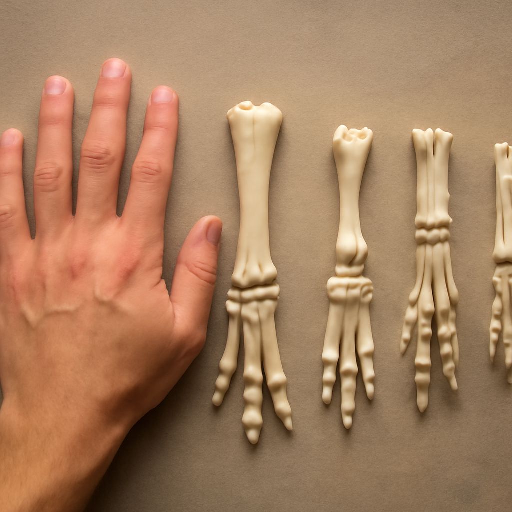
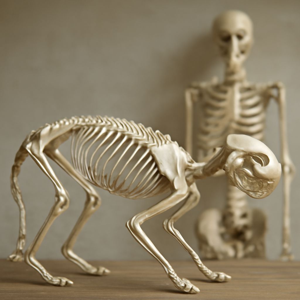
The horse skeleton contains approximately 205 bones, though this varies with lumbar vertebrae count. Horse skeleton anatomy reveals why the horse is built for speed — the long limb bones act as levers that amplify the distance covered per stride, and the fused lower limb bones reduce rotational weight at the end of each leg where it would add cost to the stride cycle.
Key landmarks in horse skeleton anatomy: the cervical vertebrae of the neck (seven bones allowing substantial head movement), the thoracic vertebrae and their attached ribs forming the barrel of the horse’s body, and the long femur of the hindlimb that corresponds to the upper leg in humans but is largely concealed by the horse’s body and hip musculature. The visible “knee” in a horse’s front leg is actually the carpus — anatomically equivalent to the human wrist.
Horse Skull Anatomy
Horse skull anatomy is dominated by the dental arcade — the horse has teeth that continue erupting throughout its life and require significant skull space to accommodate. The elongated premaxilla and maxilla bones create the characteristic long face. The orbit (eye socket) is positioned far back and high on the skull, giving horses their characteristic wide-set eyes and panoramic near-360-degree vision.
The nasal cavity in horse skull anatomy is extensive, reflecting the horse’s need to exchange large volumes of air efficiently during sustained exertion. The large, mobile ears attach to temporal and occipital skull surfaces and can rotate nearly 180 degrees independently, allowing horses to track sounds from multiple directions simultaneously.
Preparing for a Horse Anatomy Quiz
Effective preparation for a horse anatomy quiz requires active recall practice rather than passive reading. The most efficient method: cover the labels on an anatomy diagram and attempt to name each structure from memory, then check and repeat. Flashcard systems work well for terminology, but the spatial relationships between structures require diagram-based practice that forces you to locate structures relative to each other.
Focus your horse anatomy quiz preparation on the major systems tested in equine science programs: the musculoskeletal system, the respiratory system, the digestive system, and the cardiovascular system. Know both the common names and technical anatomical names for major structures, as tests often use both.
Horse Anatomy Models for Teaching
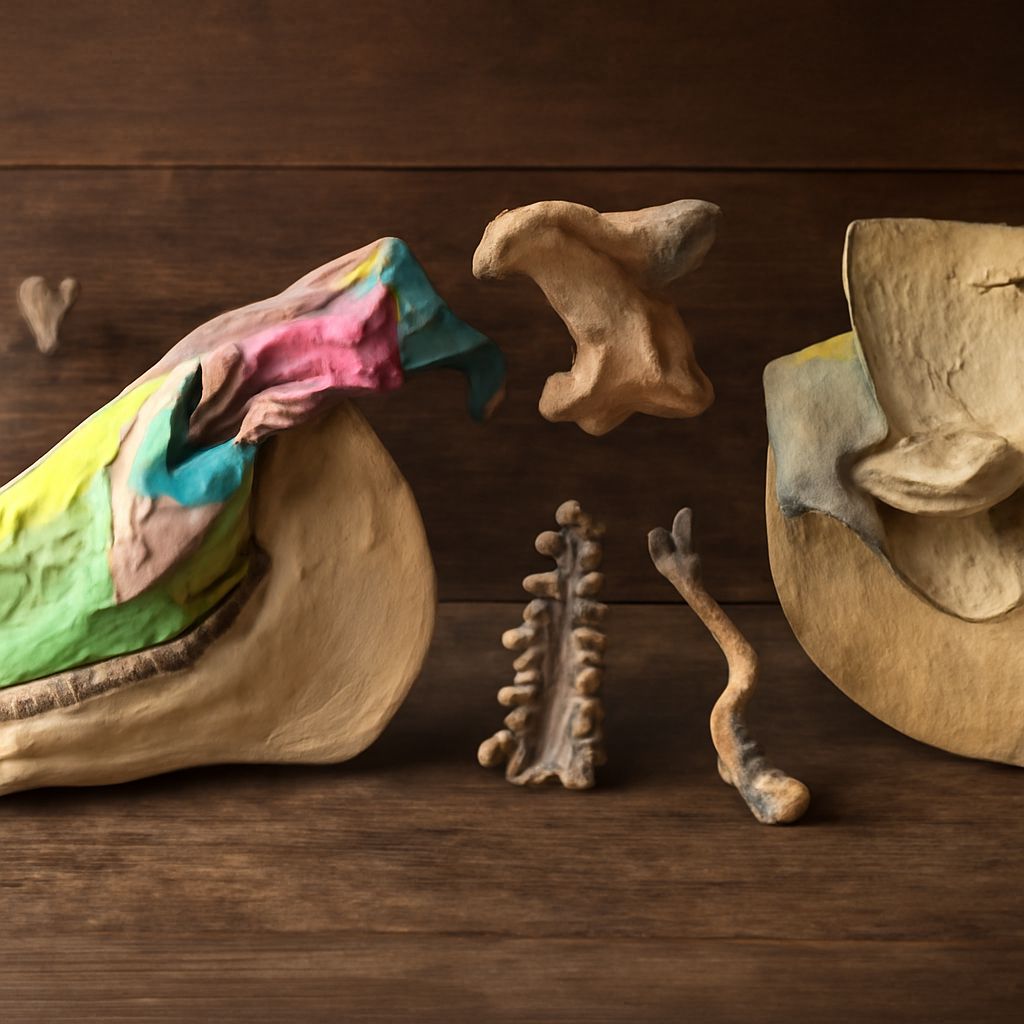
A physical horse anatomy model transforms abstract diagram study into tactile, spatial understanding. The best horse anatomy model options for educators and students include dissectible plastic models that allow you to remove and replace organ systems, painted surface anatomy models that show muscle groups without internal detail, and skeletal models with removable components for studying individual bones.
Digital anatomy tools on tablets and computers increasingly supplement physical models, allowing rotation, layer hiding, and cross-section views that physical models cannot provide. Using both physical and digital resources together produces the most complete spatial understanding of equine anatomy.