Female Muscle Anatomy: Gross Anatomy of the Muscular System Guide
Why does understanding female muscle anatomy require different emphasis than general muscular system study? The answer lies in both anatomical specifics and practical application differences. While the gross anatomy of the muscular system is largely the same between sexes — the same muscles in the same locations performing the same functions — the proportional emphasis, common training patterns, and specific regional differences in fat distribution and muscle belly shape create meaningful visual and functional distinctions worth understanding specifically. Muscle anatomy female study benefits sports scientists, fitness professionals, artists drawing the female figure, physical therapists working with female patients, and students preparing for anatomy coursework. The female muscles anatomy knowledge base connects directly to courses like exercise 13 gross anatomy of the muscular system that appears in human anatomy laboratory curricula.
This guide covers the major muscle systems with specific attention to characteristics and applications relevant to female anatomy study.
Overview of the Gross Anatomy of the Muscular System
The gross anatomy of the muscular system encompasses over 600 named skeletal muscles that attach to bone via tendons, cross at least one joint, and create movement through contraction. Studying these muscles systematically — by body region, by functional group, or by innervation — is the foundation of any anatomy curriculum. The gross anatomy of the muscular system follows consistent organizational principles: each muscle has an origin (the more stationary attachment point, usually proximal), an insertion (the more moveable attachment point, usually distal), an action (the movement produced by contraction), and innervation (the nerve that controls it).
For students in anatomy courses including exercise 13 formats, being able to identify, locate by palpation, name the origin and insertion, and describe the action of the major muscles in each region is the core competency being developed. Visual recognition from diagrams must be combined with tactile palpation on yourself and lab partners to develop the three-dimensional understanding that anatomy diagrams alone cannot provide.
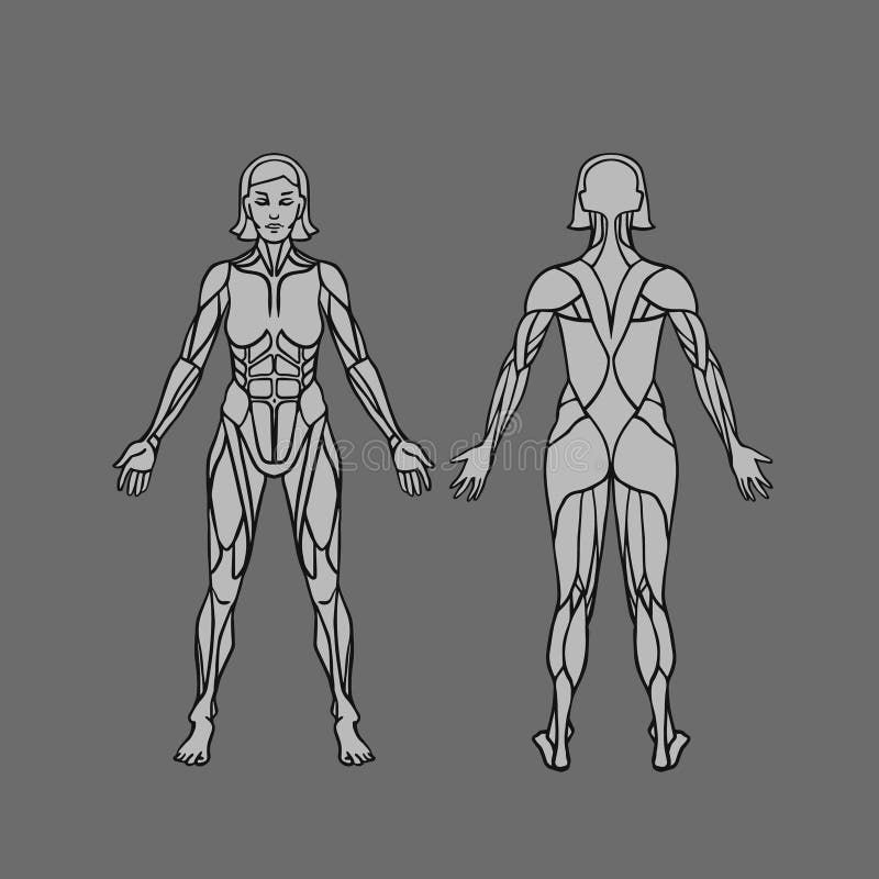
Female Muscle Anatomy: Regional Overview
The major muscle groups in female muscle anatomy follow the same regional organization as in all human anatomy — upper extremity, lower extremity, trunk, and head/neck — with consistent structure across all individuals. The muscles themselves have the same names, locations, and functions. The observable differences in female muscles anatomy are primarily in the proportions and the overlying tissue distribution rather than in muscle anatomy itself.
The gluteus maximus, medius, and minimus of the hip and buttock region are among the functionally most important muscles for female athletes and are often underemphasized in general fitness literature compared to their actual role in hip stability, knee alignment, and athletic performance. Strong gluteal muscles reduce injury risk at the knee and lower back significantly — a relationship that muscle anatomy female education in sports science and physical therapy programs emphasizes heavily.
Upper Body Muscles in Female Anatomy
Muscle anatomy female in the upper body covers the same structures as general upper body anatomy: the trapezius, rhomboids, and serratus anterior of the shoulder and back, the pectoralis major and minor of the chest, the deltoid and rotator cuff group of the shoulder, and the biceps, triceps, and forearm muscles of the arm. These muscles function identically regardless of sex.
For fitness professionals working with female clients, the posterior shoulder and upper back muscles — specifically the lower trapezius and the rhomboids — are frequently underdeveloped relative to the anterior chest and front shoulder muscles. This anterior-posterior imbalance contributes to rounded shoulders and forward head posture. Understanding female muscle anatomy in the context of common postural patterns helps trainers design corrective programming more effectively.
Core and Trunk Muscles
The core in female muscles anatomy includes the same muscles as in general anatomy: rectus abdominis, external and internal obliques, transversus abdominis, and the paraspinal erector spinae group. The transversus abdominis — the deepest layer — plays a particularly important role in spinal stability and changes significantly during pregnancy. Understanding these changes is relevant for postnatal fitness professionals and physical therapists.
The pelvic floor — a group of muscles at the base of the pelvis — is a component of female muscle anatomy that receives specific emphasis in women’s health education because of its role in continence, sexual function, and pelvic organ support. While not typically covered in standard gross anatomy courses as a primary topic, pelvic floor anatomy has significant clinical relevance in obstetrics, gynecology, and pelvic health physical therapy.
Exercise 13 Gross Anatomy of the Muscular System
In human anatomy laboratory curricula, the exercise 13 gross anatomy of the muscular system format typically covers identification of major muscles on cadaver or model specimens, mapping origins and insertions with colored pins or markers, matching muscle names to specific locations on anatomical charts, and demonstrating muscle actions through movement of articulated skeletal models. The specific content of exercise 13 gross anatomy of the muscular system varies by institution, but commonly covers the major muscles of the upper and lower limbs, the trunk and back, and the head and neck in a systematic regional approach that requires both visual identification and tactile familiarity.