Dog Leg Anatomy: A Complete Guide for Artists and Dog Owners
Do you know the difference between what you see when you look at a dog’s leg and what’s actually happening anatomically beneath the fur? Dog leg anatomy is frequently misunderstood because dogs walk on their toes (they’re digitigrade), which means the joint that looks like a dog’s “knee” bending backward on the hind leg is actually the ankle. Understanding this transforms how you interpret canine movement. Whether you’re an artist studying dog hind leg anatomy for more accurate figure drawing, a dog owner learning dog front leg anatomy for health monitoring purposes, or simply curious about the structural differences in dog rear leg anatomy, this guide covers the essential framework.
We’ll also look at how canine leg anatomy differs between front and hind limbs and why those differences matter for understanding how dogs move, bear weight, and are prone to specific injuries.
Dog Front Leg Anatomy
Bones and Joints of the Forelimb
The dog front leg anatomy starts at the scapula (shoulder blade), which floats on the rib cage connected only by muscle — dogs have no clavicle (collarbone) to lock the shoulder girdle in place the way humans do. This gives the scapula significant rotational freedom, allowing the front legs to reach forward with a longer stride than a rigid collarbone would allow.
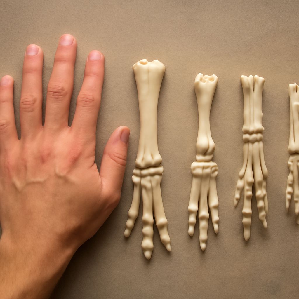
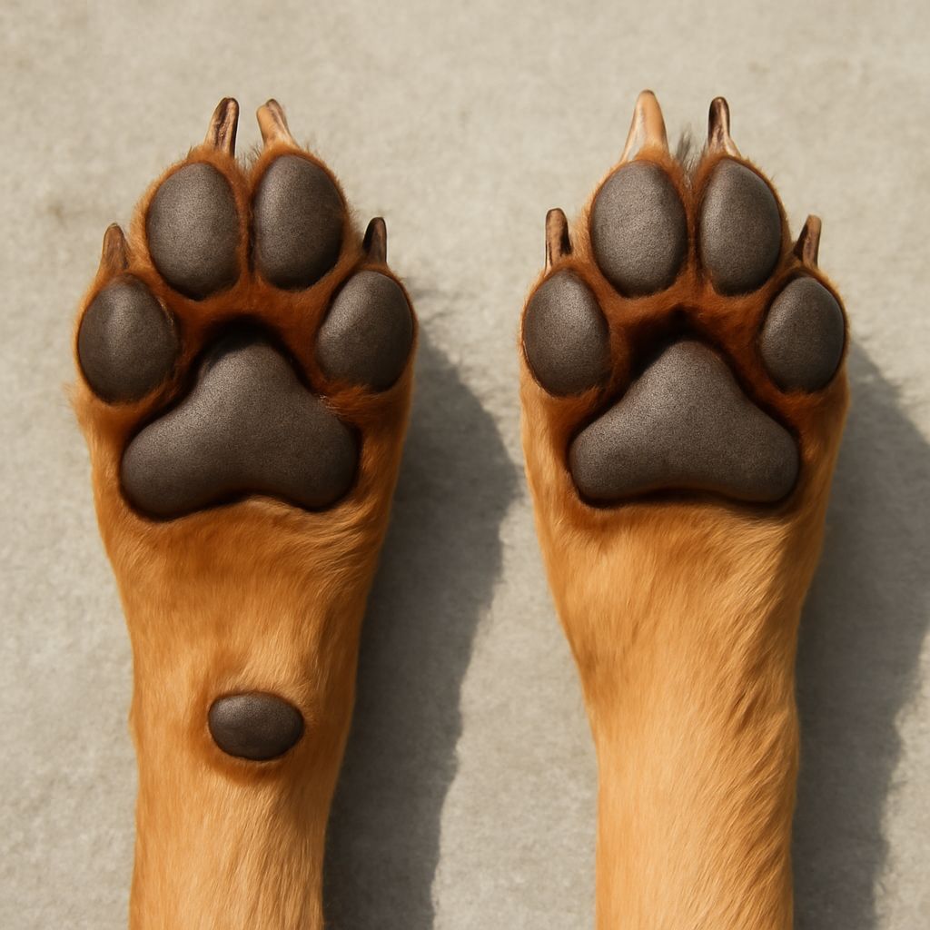
Below the shoulder is the humerus, followed by the radius and ulna (forearm bones), the carpus (the joint most people call the “wrist” — it bends forward, not backward), the metacarpal bones (equivalent to our hand bones), and the phalanges (toe bones). Dogs typically have four weight-bearing toes on each foot, plus a dewclaw on the inner aspect of the forelimb that may or may not be present depending on the breed.
Muscles of the Forelimb
The major muscles of dog front leg anatomy include the triceps (extending the elbow), the biceps brachii and brachialis (flexing the elbow), and the flexor and extensor groups of the forearm that control paw movement. The trapezius and serratus ventralis muscles connect the scapula to the spine and rib cage — they’re the suspension system that absorbs concussive force during movement and landing.
Dog Hind Leg Anatomy
The Stifle, Hock, and Paw
Dog hind leg anatomy is where the confusion between human and canine joints is most dramatic. The stifle — the joint that bends forward on the hind leg — is the true knee, equivalent to the human knee. The hock below it, which bends in the opposite direction, is the ankle. Below the hock are the metatarsal bones and the toes.
The pelvis, femur (thigh bone), stifle, tibia and fibula (shin bones), hock, metatarsals, and phalanges comprise the full chain of dog hind leg anatomy. The cruciate ligaments inside the stifle are structurally similar to the ACL and PCL in human knees — and cranial cruciate ligament (CCL) tears are among the most common orthopedic injuries in dogs, particularly in larger breeds.
Dog Rear Leg Anatomy in Motion
The hind limbs are the primary propulsion engines of canine locomotion. Understanding dog rear leg anatomy explains why dogs accelerate so powerfully from a standing start: the hip, stifle, and hock all extend simultaneously in a powerful chain of force transmission that drives the body forward. The angulation of these joints — how much they bend at rest — varies significantly by breed and directly affects athletic capacity and injury risk.
Overangulated dog rear leg anatomy (extreme bend in the stifle and hock, common in some show-bred dogs) creates a less efficient lever arm and increases stress on ligaments. More moderate angulation produces better long-term soundness for working and sporting dogs.
Canine Leg Anatomy for Artists
For artists, understanding canine leg anatomy transforms dog figure drawing from guesswork into structured observation. The key insight is the digitigrade foot posture: when you draw a dog standing, the paw is always on the ground, but the “heel” (hock on the hind leg, carpus on the front leg) is always elevated. The limb from foot to first visible joint is the equivalent of a human’s foot bones — not the lower leg.
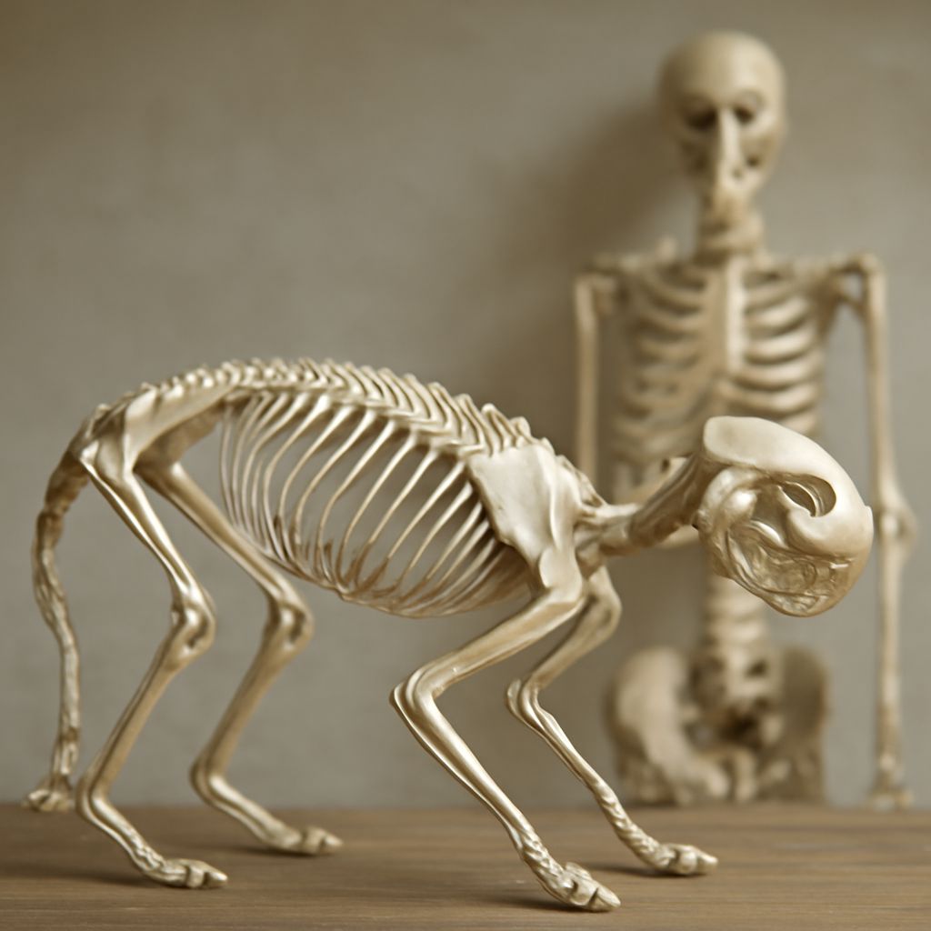
Practice drawing the skeleton of the leg before adding muscle and fur. A simple cylinder and sphere construction of each joint segment — with the correctly placed joints — produces a far more accurate result than trying to observe surface form without understanding the underlying structure. Dog leg anatomy diagrams from veterinary anatomy texts are excellent artist references precisely because they show the skeleton clearly labeled.
When drawing in motion, remember that the front legs control direction and absorb impact while the hind legs provide propulsion. This functional difference translates visually: front legs typically show more outward reach and absorptive posture while rear legs show more powerful extension. Capturing this distinction in canine figure drawing gives your dogs convincing movement energy.