Dogs Anatomy: A Complete Guide to Canine Body Systems
Have you ever looked at your dog and wondered what’s going on beneath that fur? Understanding dogs anatomy gives you a real advantage as a pet owner, helping you recognize normal structure, spot potential problems early, and communicate more clearly with your vet. From the tips of the ears to the end of the tail, every part of a dog’s body follows a logical design shaped by millions of years of evolution.
A solid grasp of canine anatomy starts with the major systems. Whether you’re consulting a dog anatomy chart for the first time or brushing up on a canine anatomy chart you studied years ago, this guide walks through each system clearly. A reliable dog anatomy reference puts the full picture together so you know exactly what you’re looking at.
Understanding the Skeletal System
Bone Count and Structure
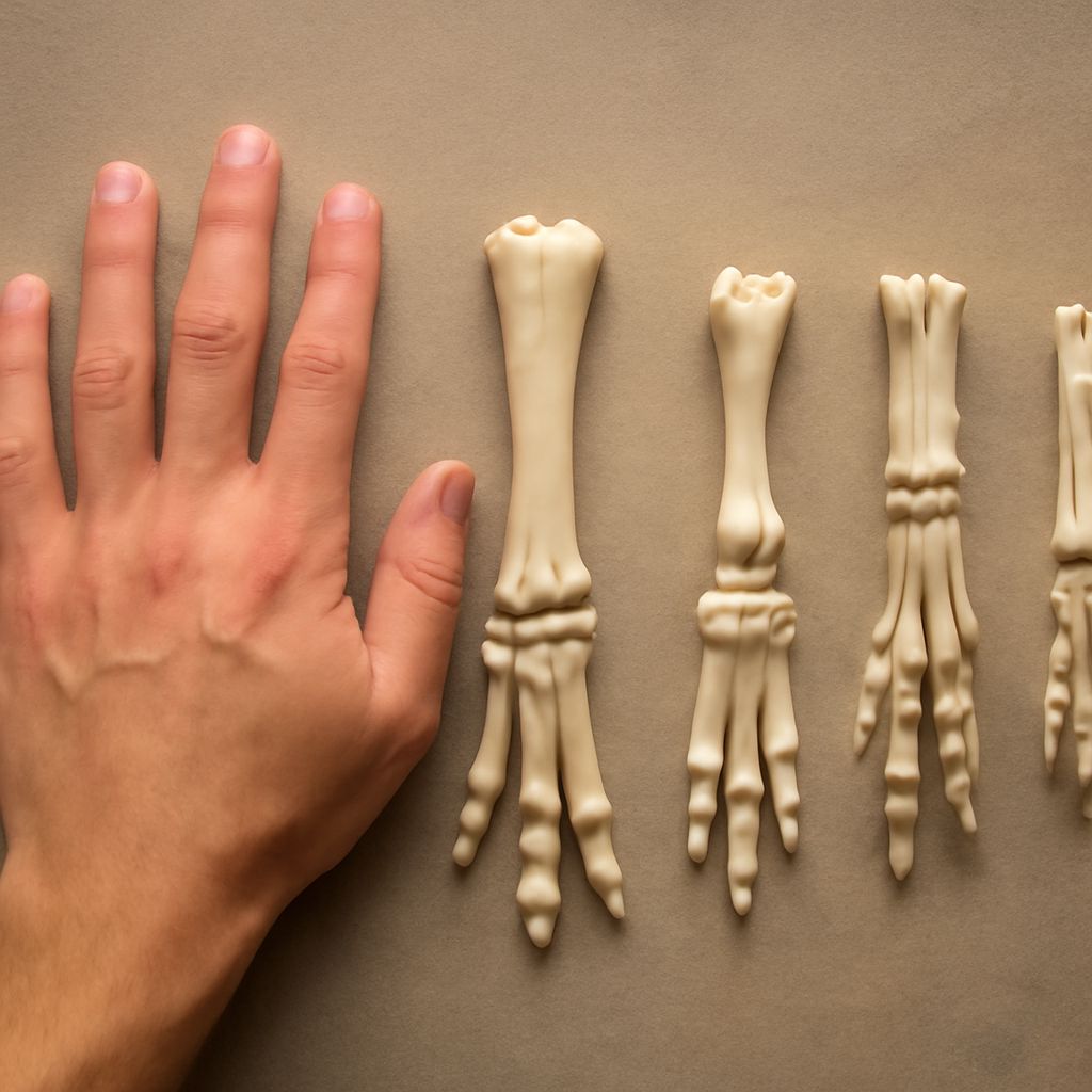
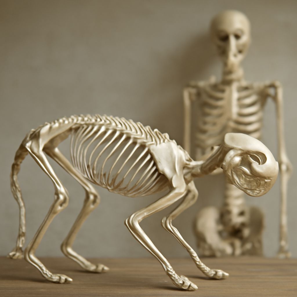
Dogs have around 319 bones, though the exact count varies slightly by breed and whether the tail was docked. The skeleton gives the body its shape, protects vital organs, and provides the leverage that makes movement possible. Compact cortical bone forms the outer shell of most bones, while spongy trabecular bone sits inside, reducing weight without sacrificing strength.
The Spine and Limbs
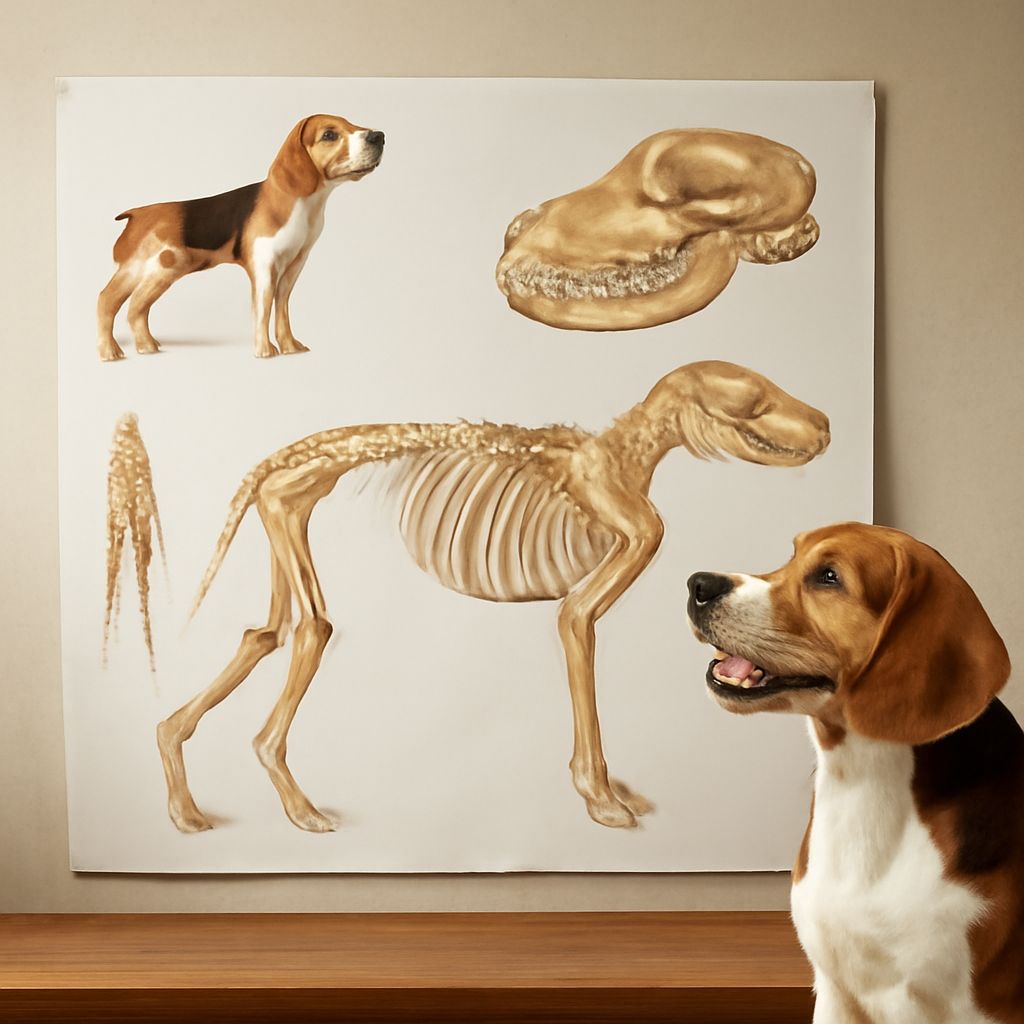
The vertebral column runs from the skull to the tail tip and divides into cervical, thoracic, lumbar, sacral, and caudal sections. The front limbs attach to the body through the scapula and a muscular sling rather than a true ball-and-socket joint, which gives dogs a wider range of motion for digging and climbing. Rear limbs connect at the hip joint and drive the powerful push-off that fuels running gaits. Any dogs anatomy guide worth reading will highlight how the angle of the stifle (knee) affects speed and agility.
Skull and Jaw Mechanics
Skull shape varies enormously across breeds. Brachycephalic breeds like bulldogs have shortened facial bones that alter breathing pathways, while dolichocephalic breeds like greyhounds have elongated skulls built for wide-angle vision. The temporomandibular joint allows the jaw to open wide, and the carnassial teeth — the large shearing premolars — are a defining feature of canine dental anatomy.
Muscular and Soft Tissue Layout
Major Muscle Groups
The epaxial muscles run along the top of the spine, stabilizing posture and extending the back. Hypaxial muscles on the underside flex the spine and protect the abdomen. The gluteal and hamstring muscles of the rear quarters generate the most raw power during acceleration. In the forelimb, the brachiocephalicus and triceps work together to extend and retract the leg through each stride. Any canine anatomy chart labels these groups clearly, helping you visualize how force moves through the body.
Tendons and Ligaments
Tendons connect muscle to bone and transmit the pull of each contraction into movement. Ligaments bind bone to bone and limit range of motion to safe angles. The cruciate ligaments inside the stifle joint are among the most commonly injured in active dogs, especially larger breeds. Understanding where these soft tissues sit — something a dog anatomy reference makes visual and immediate — helps you recognize signs of strain before an injury becomes serious.
Internal Organs and Systems
Digestive Tract Overview
Dogs are monogastric omnivores with a digestive system optimized for processing both animal protein and plant material. Food travels from the mouth through the esophagus, into the stomach — which can expand dramatically — and then through the small intestine where most nutrient absorption occurs. The large intestine reabsorbs water and forms waste. Gastric dilatation-volvulus, or bloat, is a life-threatening condition where the stomach twists, cutting off circulation. Knowing basic dogs anatomy of the digestive tract helps you understand why this condition progresses so quickly.
Respiratory and Cardiovascular Systems
The canine respiratory system draws air through the nose or mouth, past the larynx, down the trachea, and into two lungs divided into lobes. Dogs regulate body temperature primarily through panting, not sweating. The heart sits in the chest between the lungs, with the left side pumping oxygenated blood to the body and the right side routing deoxygenated blood to the lungs. Breed-specific heart conditions, like dilated cardiomyopathy in large breeds, are easier to monitor when you understand normal canine anatomy.
Reproductive Anatomy Basics
Intact females have a bicornuate uterus with two uterine horns, which allows for large litters. Males have testes that typically descend into the scrotum by eight weeks of age. Cryptorchidism — when one or both testes fail to descend — is a common issue identified during routine examination. A thorough dog anatomy reference will include both male and female reproductive layouts for complete understanding.
Reading a Dog Anatomy Chart
How to Use Reference Diagrams
A dog anatomy chart works best when you use it actively. Cross-reference the labeled regions with what you can feel on your own dog during a calm grooming session. You’ll quickly learn to locate the iliac crest, the olecranon, and the costal arch by touch. A canine anatomy chart that shows both skeletal and muscular overlays gives you a layered understanding that flat diagrams alone cannot provide.
Common Chart Annotations
Most charts use directional terms borrowed from human anatomy, adjusted for quadruped orientation. Cranial means toward the head, caudal means toward the tail, dorsal refers to the back, and ventral to the belly. Medial and lateral describe position relative to the midline. Learning these terms makes any dog anatomy reference far more useful, because you can read veterinary notes and ask informed questions during appointments.