Knee Anatomy Pain, Dog Tail Anatomy, and Canine Body Structure Explained
Do you ever wonder why knee anatomy pain is such a common complaint in both humans and their canine companions? Or how dog tail anatomy relates to your pet’s emotional communication and physical health? Understanding the structural foundations of bodies — whether human knees or a dog’s complex musculoskeletal system — helps you recognize normal function, spot potential problems early, and engage more meaningfully with veterinary or medical guidance. This guide covers knee anatomy pain in context, then shifts focus to dog tail anatomy, dog mouth anatomy, dog knee anatomy, and visual dog infographic references for owners who want to learn through images.
Whether you’re an artist studying animal anatomy, a dog owner concerned about your pet’s health, or a student preparing for anatomy coursework, you’ll find clear, organized information ahead. Let’s start with the basics and build toward a comprehensive picture.
Understanding Knee Anatomy Pain
The Knee Joint Structure
Knee anatomy pain most commonly originates from one of four key structures: the menisci (cartilage cushions), the cruciate ligaments, the patellar tendon, or the articular cartilage surfaces of the femur and tibia. The knee is a modified hinge joint designed to handle enormous forces — up to six times body weight during running — making it inherently vulnerable to both acute injury and chronic wear.
Understanding the source of knee anatomy pain requires knowing which structure is involved. Medial knee pain often implicates the medial meniscus or medial collateral ligament; pain behind the kneecap suggests patellar tracking issues; deep joint pain during weight-bearing activities may indicate articular cartilage degradation. Always consult a qualified healthcare provider for diagnosis and treatment of persistent joint discomfort.
Dog Tail Anatomy
Structure and Function of the Tail
Dog tail anatomy is built around a series of small caudal vertebrae — typically six to twenty-three bones, depending on the breed. These vertebrae are held together by facet joints and surrounded by muscles, tendons, and nerves that control tail movement. The tail connects to the sacrum at the base of the spine through the sacrocaudal junction, a region that can be injured during rough play or accidental trauma.
Understanding dog tail anatomy goes beyond curiosity — it explains why tail injuries are more serious than they might appear. Damage to the caudal vertebrae or the surrounding nerve structures can affect bladder and bowel control if the injury is close enough to the sacral plexus. A limp or painful tail warrants veterinary evaluation rather than a wait-and-see approach.
Dog Mouth Anatomy
Dog mouth anatomy differs significantly from human oral structure in several important ways. Dogs have 42 adult teeth (humans have 32), including carnassial teeth — the large shearing molars and premolars designed for processing meat and bone. The jaw mechanics of dog mouth anatomy allow for powerful vertical crushing force but limited lateral grinding motion, unlike the side-to-side chewing action humans use.
The tongue in dog mouth anatomy is longer and more mobile than a human tongue, serving crucial roles in thermoregulation (panting), grooming, and food manipulation. The salivary glands produce large quantities of saliva with strong antibacterial properties, which is part of why dogs instinctively lick wounds.
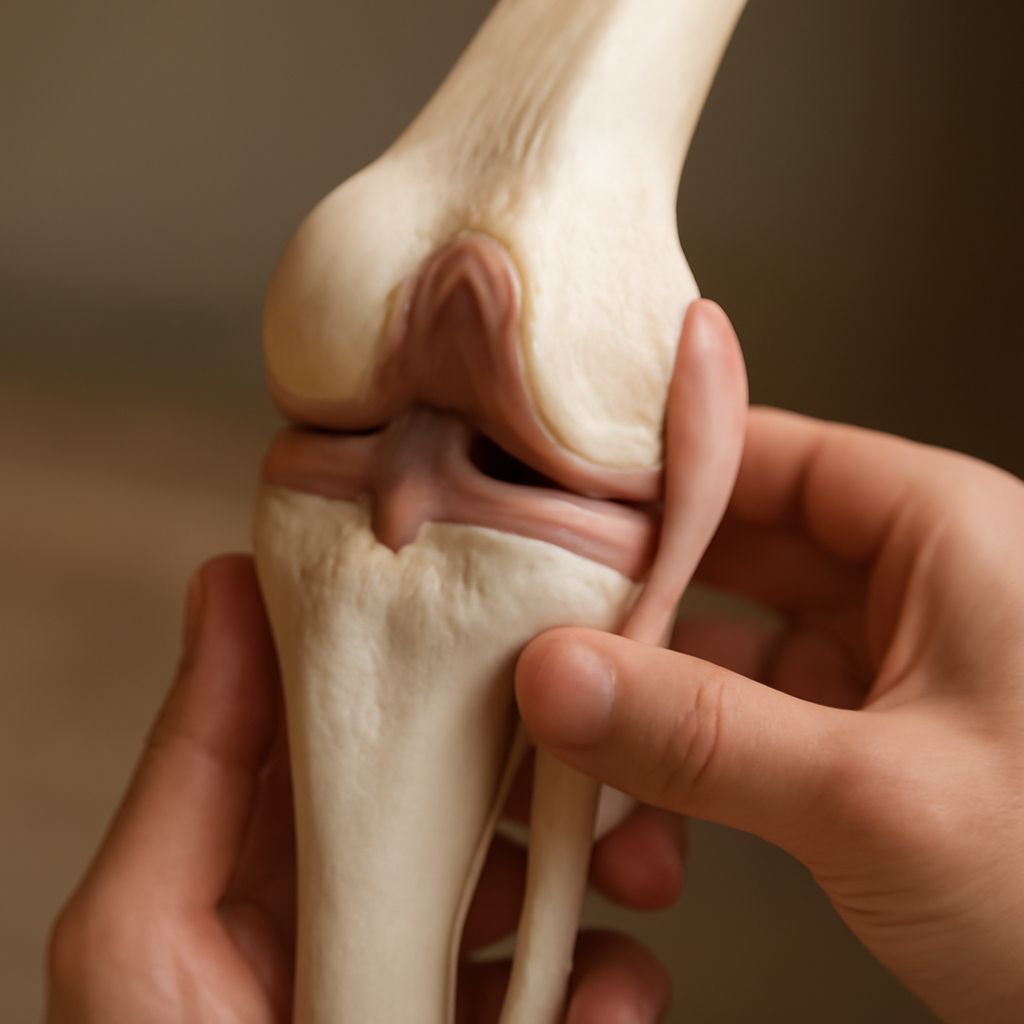
Dog Knee Anatomy
Dog knee anatomy centers on the stifle joint — the canine equivalent of the human knee — which connects the femur, tibia, and fibula. The stifle contains two cruciate ligaments (cranial and caudal), two menisci, and multiple supporting ligaments that stabilize the joint during movement. Cranial cruciate ligament (CCL) rupture is one of the most common orthopedic injuries in dogs, particularly in large breeds, closely paralleling the ACL injuries familiar in human athletes.
Recognizing signs of dog knee anatomy problems — a sudden hind-leg limp, reluctance to bear weight, or a popping sensation in the joint — allows you to seek veterinary care before secondary damage occurs. Surgical intervention and controlled rehabilitation are the standard treatment approaches for significant stifle injuries.
Using a Dog Infographic for Learning
A well-designed dog infographic simplifies complex anatomical information into a visual format that’s easy to absorb and remember. Quality dog infographic resources label the major skeletal and muscular structures, highlight common injury sites, and often include comparison charts between dog and human anatomy. Veterinary educational publishers, professional associations, and online platforms like Veterinary Partner publish peer-reviewed dog infographic materials suitable for both owners and students. Bookmark a reliable dog infographic reference alongside this text for a complete learning toolkit.