Knee Anatomy Ligaments: Understanding the Complex Structure
How well do you understand the anatomy of a knee? If you’ve ever experienced knee pain or injury, you know how crucial it is to understand knee ligament anatomy. The knee is a complex joint, one of the largest in the human body, and it’s held together by an intricate network of ligaments. But what roles do these ligaments play in the anatomy of a knee, and how can you visualize and maintain them effectively? This comprehensive guide delves into the essential aspects of knee ligaments anatomy, exploring everything from the types and functions of each ligament to practical tips for their care and maintenance.
Introduction to the Anatomy of a Knee
Overview of Knee Structure
The knee is a pivotal hinge joint that connects the thigh bone (femur) to the shin bone (tibia). In addition to these bones, the fibula, a smaller bone running alongside the tibia, and the kneecap (patella) are key components of the knee structure. The knee is equipped with cartilage that cushions and absorbs shock, as well as synovial fluid to reduce friction during movement. However, it’s the knee ligaments anatomy that plays a critical role in stabilizing this joint.
Importance of Knee Ligaments
Knee ligaments are strong bands of tissue that connect bones and provide stability to the knee joint. Each ligament has a specific function, contributing to the overall mobility and support of the knee. Understanding knee ligament anatomy is crucial for anyone looking to maintain healthy, functional knees. It helps you recognize the importance of these structures in everyday activities and the impact of injuries on your mobility.
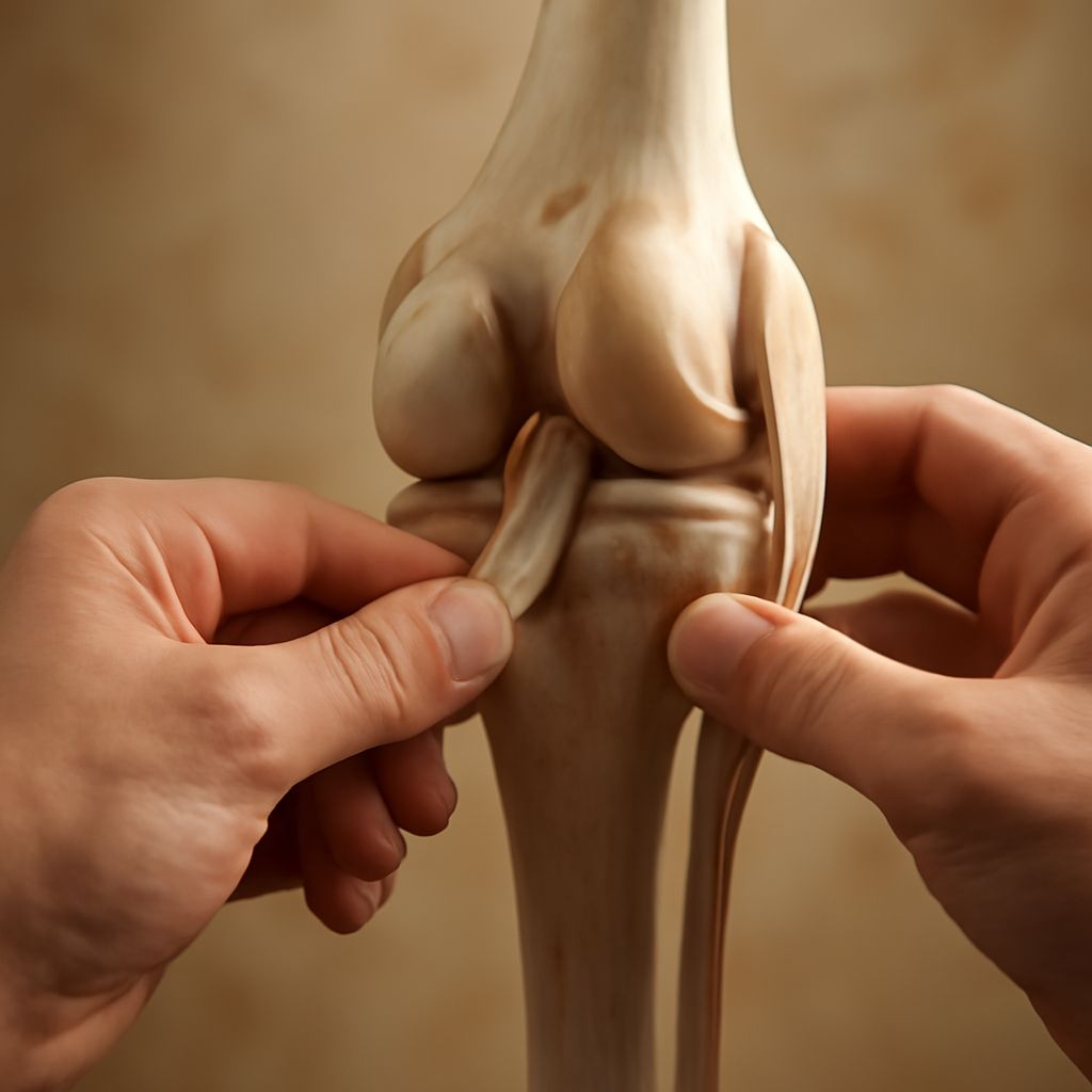
Detailed Knee Ligament Anatomy
Types of Knee Ligaments
There are four primary ligaments that form the core of the knee’s stability: the anterior cruciate ligament (ACL), posterior cruciate ligament (PCL), medial collateral ligament (MCL), and lateral collateral ligament (LCL). Each plays a unique role in the knee ligaments anatomy. The ACL and PCL cross each other inside the knee, controlling the back-and-forth motion, while the MCL and LCL are located on either side of the knee, managing side-to-side movement.
Functions of Each Ligament
The ACL prevents the tibia from sliding out in front of the femur, a crucial function for activities that involve stopping and changing direction. The PCL, on the other hand, prevents the tibia from sliding backwards under the femur. The MCL resists forces pushing the knee inward, and the LCL counteracts forces pushing the knee outward. Each ligament’s specific function is integral to the stability and performance of the knee joint.
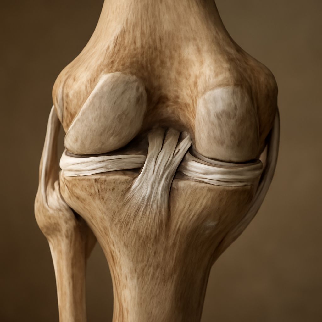
Visualizing Knee Ligaments Anatomy
Knee Anatomy Images and Diagrams
Visual aids can significantly enhance your understanding of knee ligaments anatomy. Knee anatomy images and diagrams provide detailed views of the ligaments and other components of the knee. They illustrate how each ligament is positioned and how they interact with bones and cartilage. These images are particularly useful for identifying the exact location of ligaments and understanding their spatial relationships within the joint.
Tips for Understanding Images
When examining knee anatomy images, focus on both the overall structure and individual components. Look for labeled diagrams that clearly mark each ligament, bone, and cartilage. These illustrations help you connect two-dimensional images to the three-dimensional structure of a real knee. Utilize these visuals to gain a better grasp of knee ligament anatomy and how it contributes to joint function.
Maintaining Healthy Knee Ligaments
Common Knee Injuries
Knee injuries are common, especially among athletes and active individuals. The most frequent injuries involve the ACL, often resulting from sudden changes in direction or improper landings. The MCL and LCL can also be injured due to direct impact or twisting motions. Understanding knee ligaments anatomy helps you identify potential risks and take necessary precautions to avoid such injuries.
Prevention and Care Tips
Maintaining healthy knee ligaments requires a combination of proper exercise, stretching, and protective measures. Regular strengthening exercises for the muscles around the knee, including the quadriceps and hamstrings, support the ligaments and enhance joint stability. Flexibility exercises, such as stretching, help maintain ligament elasticity. Additionally, using appropriate footwear and protective gear during high-impact activities is essential. By understanding the knee ligament anatomy and adhering to these preventative measures, you can reduce the risk of injury and preserve the health of your knees.