Cat Skeleton Anatomy: A Complete Guide to Feline Bones and Structure
Have you ever watched a cat squeeze through a gap that seems impossibly small and wondered how its body manages that feat? The answer lies in cat skeleton anatomy — a remarkably flexible framework built for agility, speed, and precision. Understanding cat leg anatomy, including the subtle details of cat front leg anatomy, helps artists render felines accurately and gives pet owners insight into how their companions move. Add in cat anatomy bones knowledge and a look at big cat anatomy, and you have a full picture of one of nature’s most efficient body plans.
You don’t need a biology degree to appreciate feline skeletal structure. A working knowledge of key bones and their roles transforms your ability to sketch cats, assess posture, or simply marvel at how they operate. This guide walks you through every major region, from skull to tail tip.
Overview of Cat Skeleton Anatomy
The Axial Skeleton
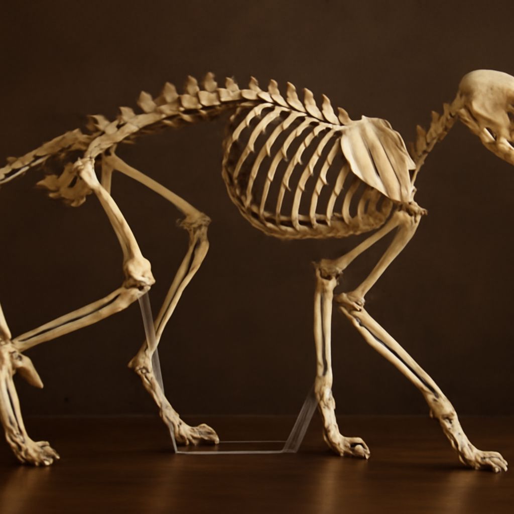
Cat skeleton anatomy begins with the axial skeleton — the skull, vertebral column, and ribcage. Cats have around 230 bones, slightly more than humans, largely because of their long, mobile tails. The vertebrae are loosely connected, allowing the spine to twist and arch far beyond what most mammals manage. This hypermobility is why cats land on their feet: the spine rotates independently of the pelvis in freefall.
The Appendicular Skeleton
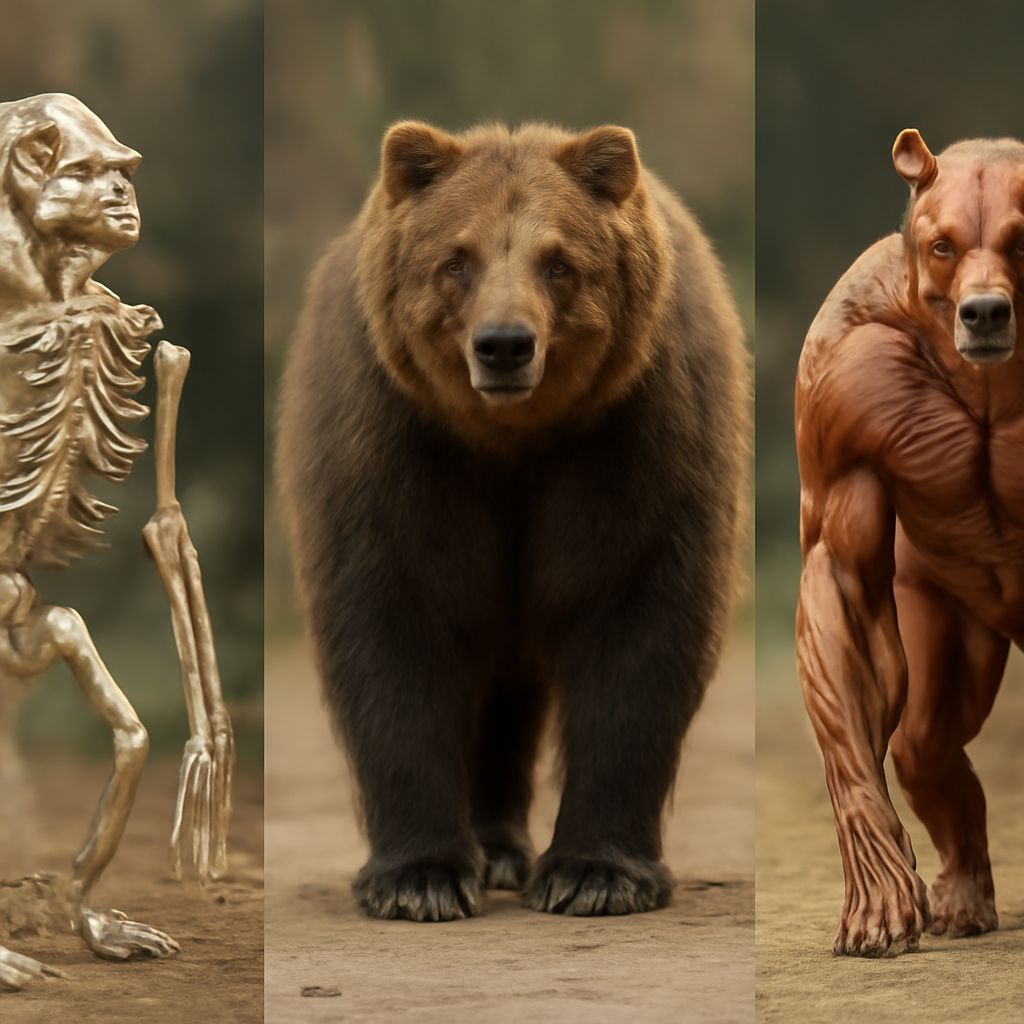
The appendicular portion covers the limbs and their girdles. The clavicle in cats is vestigial — just a small floating bone — which removes the rigid shoulder lock found in humans and lets the front legs rotate freely. This skeletal freedom makes cats excellent climbers and leapers.
Unique Feline Skeletal Adaptations
Cats walk digitigrade — on their toes, not their full foot. This elongates the effective leg length without adding height, giving each stride extra reach. The lightweight skull and forward-facing eye sockets reflect a predator design optimized for depth perception and fast bite mechanics.
Cat Leg Anatomy: Front and Rear Limbs
Cat Front Leg Anatomy in Detail
Cat front leg anatomy includes the scapula, humerus, radius, ulna, carpals, metacarpals, and phalanges. The scapula sits loosely against the ribcage, cushioning impacts during landing. When you study cat front leg anatomy closely, you notice that the radius and ulna allow a degree of rotation that helps the cat swipe at prey or manage rough terrain. The wrist sits high off the ground compared to the knuckle-level wrist of a dog.
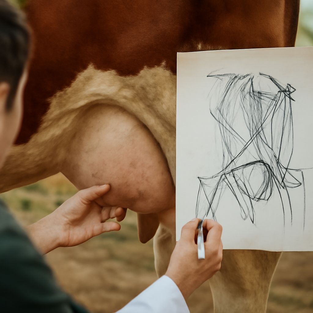
Artists often misplace the elbow when drawing cats because it sits closer to the body than expected. Reference the humerus length — roughly equal to the distance from the elbow to the wrist — to place limbs accurately.
Rear Leg Structure and Function
Cat leg anatomy in the rear features the femur, tibia, fibula, tarsals, and metatarsals. The hock — which looks like a backward knee — is actually the ankle. Long metatarsals act as a spring-loaded lever, storing and releasing energy with each stride. This design gives cats explosive acceleration from a standing start.
How Limb Angles Power Movement
The angle between femur and tibia in a resting cat is around 100 degrees — a coiled spring ready to extend. When a cat launches into a jump, that angle opens rapidly, converting stored muscular tension into vertical height. Understanding these angles sharpens both your anatomical drawings and your appreciation of feline athleticism.
Cat Anatomy Bones: Key Landmarks
Skull and Jaw Bones
Cat anatomy bones in the skull include the frontal, parietal, temporal, and occipital bones forming the cranium, plus the zygomatic arch that creates the wide cheekbone profile. The lower jaw, or mandible, carries large canine teeth designed to grip and hold prey. The temporomandibular joint allows the jaw to open wide but does not slide sideways — cats tear rather than grind.
Spine and Ribcage
Among cat anatomy bones, the 13 thoracic vertebrae each attach to a rib, forming a deep but narrow ribcage. This shape lets the cat compress its body horizontally to fit through tight spaces. The lumbar vertebrae — usually seven — are especially flexible and thick, supporting the muscular hindquarters that drive sprinting power.
Paws and Claws
The distal phalanges of each digit carry retractable claws in most feline species. A ligament holds the claw retracted at rest; contraction of flexor muscles extends it on demand. Cheetahs are a notable exception — their claws are semi-retractable and act as cleats for traction at high speed.
Big Cat Anatomy Compared to Domestic Cats
Skeletal Scale Differences
Big cat anatomy follows the same blueprint as domestic cats but with significant proportional shifts. Lions and tigers carry heavier skulls with more pronounced sagittal crests for anchoring powerful jaw muscles. The limb bones are denser and the overall frame is broader to support substantial body mass. A tiger’s femur can be four times longer than a house cat’s while maintaining nearly identical joint angles.
Hunting Adaptations in Large Felines
Big cat anatomy reflects each species’ hunting strategy. The cheetah has a flexible spine that acts like a spring, extending stride length beyond what leg length alone could achieve. Leopards carry large shoulder muscles and a wide scapular range of motion to haul prey into trees. Jaguars have the heaviest skull-to-body ratio among big cats, generating a bite force that can pierce turtle shells.
What Artists Can Learn from Big Cats
Studying big cat anatomy refines your domestic cat drawings. The exaggerated proportions — longer limbs, broader shoulders, more defined muscle over bone — make structural landmarks easier to identify and then scale down. If you can draw a lion skeleton convincingly, rendering a housecat’s subtler framework becomes intuitive. Overlay reference images of both to practice proportion scaling.