Hand Tendon Anatomy, Thumb and Peroneal Tendons Explained
Have you ever wondered what’s actually happening beneath the skin when you flex your fingers, extend your thumb, or point your foot? Hand tendon anatomy is a remarkable story of precise engineering — dozens of tendons working in coordinated harmony to transmit the force of forearm and leg muscles to the small bones of the hand, thumb, and ankle. Understanding anatomy of the hand tendons, along with thumb tendon anatomy and peroneal tendon anatomy in the ankle, gives you a functional map of structures that are commonly injured, commonly misunderstood, and critically important to everyday movement.
This guide also touches on a different kind of “anatomy” — the anatomy of the constitution — to explore how systematic structural analysis serves both biological and legal understanding. Whether you’re a student, a healthcare practitioner, or a curious reader, you’ll find clear, organized information throughout.
Hand Tendon Anatomy: Flexors and Extensors
Flexor Tendon System
Hand tendon anatomy begins with the two major flexor tendons that serve each finger (except the thumb): the flexor digitorum superficialis (FDS) and the flexor digitorum profundus (FDP). The FDS tendon inserts on the middle phalanx and bends the proximal interphalangeal (PIP) joint. The FDP tendon passes through a split in the FDS tendon and inserts on the distal phalanx, bending the distal interphalangeal (DIP) joint — the outermost knuckle — as well as contributing to PIP flexion.
These tendons travel through fibrous sheaths called tendon sheaths, held close to the bone by a series of annular and cruciate pulleys that prevent bowstringing. Inflammation of these sheaths (tenosynovitis) causes trigger finger; rupture of the A1 pulley is common in rock climbers. Anatomy of the hand tendons includes this pulley system as an essential functional component, not merely the tendons themselves.
Extensor Tendon System
The extensor tendons of hand tendon anatomy travel along the dorsal (back) surface of the hand and fingers. They’re organized into a complex structure called the extensor mechanism or extensor hood at the finger level — a fan of tendon fibers that spreads across the proximal phalanx and splits into three bands: a central slip inserting on the middle phalanx, and two lateral bands that recombine over the middle phalanx to insert on the distal phalanx. This elegant arrangement allows the extensor tendons to straighten all three finger joints from a single muscle system.
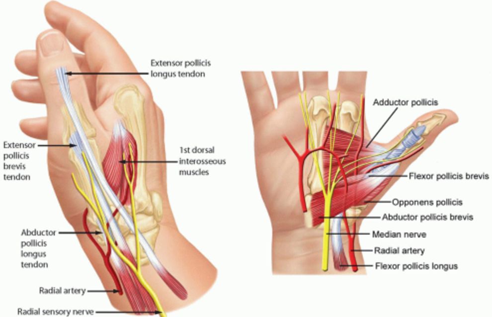
Thumb Tendon Anatomy
Thumb tendon anatomy differs from the other digits because the thumb has only two phalanges and its own dedicated set of tendons. The flexor pollicis longus (FPL) is the sole flexor tendon serving the thumb’s interphalangeal joint, running through a distinct tendon sheath on the thumb’s palmar surface. Two extensor tendons serve the thumb: the extensor pollicis longus (EPL) extends the IP joint and passes around Lister’s tubercle on the radius; the extensor pollicis brevis (EPB) extends the MCP joint.
Understanding thumb tendon anatomy is clinically important because EPL rupture — sometimes occurring after wrist fractures or rheumatoid arthritis — produces an inability to extend the thumb’s tip, a condition that significantly impairs grip and pinch function. The distinctive “anatomical snuffbox” visible on the dorsum of the wrist is bounded by the EPB and abductor pollicis longus tendons anteriorly and the EPL tendon posteriorly — a landmark used in clinical assessment of scaphoid fractures.
Peroneal Tendon Anatomy
Peroneal tendon anatomy moves from the hand to the ankle region, where two peroneal tendons — the peroneus longus and peroneus brevis — travel behind the lateral malleolus (the bony prominence on the outside of the ankle) and contribute to foot eversion and plantar flexion. The peroneus brevis inserts on the base of the fifth metatarsal; the peroneus longus crosses under the foot to insert on the medial cuneiform and first metatarsal, providing an important stabilizing arch support function.
Peroneal tendon anatomy is clinically significant because peroneal tendon tears, subluxation, and tenosynovitis are common in athletes who participate in sports involving lateral movements, ankle sprains, or repetitive cutting actions. Understanding the anatomical course of the peroneal tendons behind and below the lateral malleolus helps explain the mechanism of these injuries and guides appropriate treatment decisions.
The Anatomy of the Constitution
The phrase “anatomy of the constitution” describes the structural analysis of constitutional documents — dissecting their components, understanding how each element functions, and tracing the relationships between provisions. Just as hand tendon anatomy maps functional structures within the hand, constitutional anatomy maps the foundational structures of democratic governance: the distribution of powers, the system of checks and balances, the amendment process, and the rights guarantees.