Thumb Anatomy: Hand Muscles, Nerves, and Complete Thumb Structure Guide
Have you ever stopped to appreciate how much of your daily function depends on your thumb? Thumb anatomy is uniquely complex among the digits — it’s the only finger that can oppose the others, enabling the precision grip that defines much of human tool use and fine motor skill. Understanding hand muscles anatomy in relation to the thumb, along with the anatomy of thumb joints and the critical hand anatomy nerves that make sensation and control possible, gives you a deeper appreciation of this remarkable structure.
Whether you’re a healthcare student, an occupational therapist, an artist learning to draw hands accurately, or someone recovering from a thumb injury, this guide provides clear, organized information. We’ll cover the anatomy of the thumb from bone structure through muscle groups to nerve supply, helping you understand how everything works together.
Bones of the Thumb
The Two-Phalanx Structure
Unlike the other four fingers, which each have three phalanges, thumb anatomy features only two: the proximal phalanx and the distal phalanx. This two-bone structure is part of what enables the thumb’s unique range of motion. The proximal phalanx connects to the first metacarpal at the metacarpophalangeal (MCP) joint — the main knuckle of the thumb — while the distal phalanx connects to the proximal phalanx at the interphalangeal (IP) joint.
The first metacarpal, which forms the base of the anatomy of thumb structure, articulates with the trapezium bone of the wrist at the carpometacarpal (CMC) joint — the saddle joint that allows the thumb’s signature opposition movement. Damage to this CMC joint (the most common site of thumb osteoarthritis) dramatically impairs pinch grip and daily function.
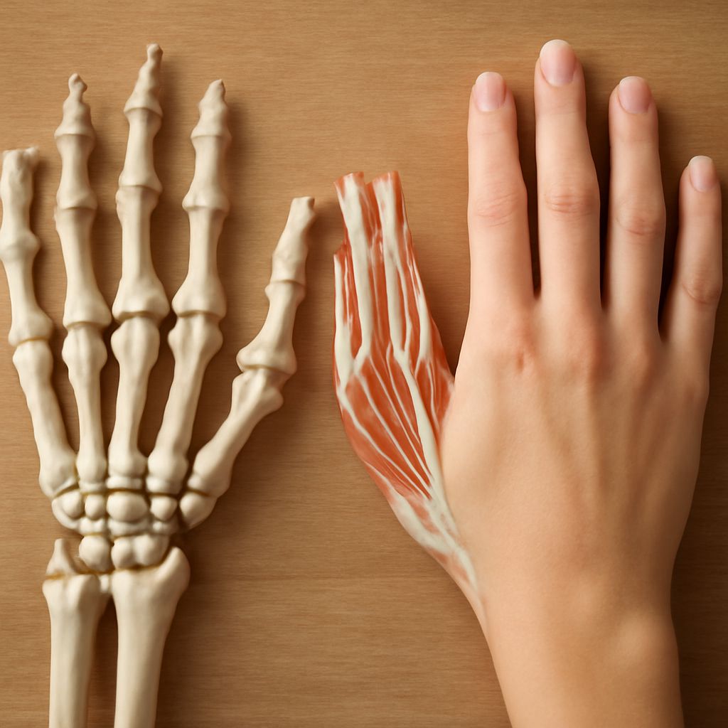
Hand Muscles Anatomy: The Thenar Group
The thenar eminence — the muscular pad at the base of your thumb — is the primary expression of hand muscles anatomy related to thumb function. Four intrinsic muscles form the thenar group: the abductor pollicis brevis, flexor pollicis brevis, opponens pollicis, and adductor pollicis. Together, these muscles enable the full range of thumb movements: abduction (moving away from the index finger), flexion (bending), opposition (rotating to face the other fingers), and adduction (returning to the resting position alongside the index finger).
The anatomy of the thumb also depends on extrinsic muscles originating in the forearm: the flexor pollicis longus flexes the thumb’s IP joint, while the extensor pollicis longus and brevis extend the IP and MCP joints respectively. The abductor pollicis longus contributes to radial abduction and extension. Understanding this interplay of hand muscles anatomy — intrinsic thenar muscles working alongside extrinsic forearm muscles — explains why both hand and forearm injuries can impair thumb function.
Anatomy of the Thumb: Ligaments and Stability
The CMC saddle joint’s mobility comes at the cost of inherent stability, making the anatomy of thumb ligamentous structures critically important. Several strong ligaments surround the trapezio-metacarpal joint, preventing excessive translation while allowing the broad arc of movement. The ulnar collateral ligament (UCL) of the MCP joint is the most clinically relevant structure here — UCL tears (commonly called “skier’s thumb” or “gamekeeper’s thumb”) produce painful instability of the main knuckle and require specific treatment.
Hand Anatomy Nerves Supplying the Thumb
Three nerves contribute to hand anatomy nerves supply of the thumb. The median nerve supplies sensation to the palmar (volar) surface of the thumb and the radial two-thirds of the palm — critical for the fine-touch discrimination needed for precision grip. The radial nerve supplies the dorsal (back) surface of the thumb and the first web space. The ulnar nerve contributes motor supply to the adductor pollicis and part of the flexor pollicis brevis.
Carpal tunnel syndrome — compression of the median nerve at the wrist — specifically impairs the hand anatomy nerves serving the thumb’s palmar sensation and the thenar muscles, explaining why patients report weakness in pinching and difficulty with fine motor tasks. Understanding the nerve supply to the anatomy of the thumb helps you interpret these clinical patterns and appreciate why different nerve injuries produce different functional deficits.
Key takeaways: Thumb anatomy is distinguished by its two-phalanx structure, unique saddle joint at the CMC level, and four-muscle thenar group that enables opposition. Hand muscles anatomy in the thenar eminence works alongside extrinsic forearm muscles for full thumb function. Always consult a qualified healthcare provider for specific diagnosis and treatment of thumb injuries or symptoms.