Shoulder Anatomy: A Complete Guide to Bones, Muscles, and Joints
Have you ever wondered why the shoulder can move in so many directions while still holding your arm in place? Shoulder anatomy is one of the most intricate arrangements in the human body, built for both stability and a wide range of motion. Understanding the anatomy of shoulder structures helps you make sense of why this joint is so capable and, at times, so vulnerable to injury.
From the bones that form the framework to the muscles that drive every lift and rotation, human shoulder anatomy involves multiple overlapping systems working together. Whether you are recovering from an injury, studying for an exam, or simply curious, learning about anatomy shoulder organization gives you practical insight into how your upper body actually works.
Key Bones in Shoulder Anatomy
The Clavicle and Its Role
The clavicle, commonly called the collarbone, runs horizontally from the sternum to the tip of the shoulder. In shoulder anatomy, it acts as a strut that keeps the shoulder blade positioned away from the chest wall. Without it, the entire shoulder would collapse inward. The clavicle is the most commonly broken bone in the upper body, which says a lot about the forces it routinely absorbs.
The Scapula Explained
The scapula, or shoulder blade, is a flat triangular bone that sits against the back of the rib cage. It provides attachment points for more than a dozen muscles and forms part of two key shoulder joints. The acromion, a bony projection at the top of the scapula, is what you feel at the highest point of your shoulder.
The Humerus: Top of the Arm
The humerus is the long bone of the upper arm. Its rounded head fits into the glenoid cavity of the scapula, forming the main ball-and-socket joint of the shoulder. The shape of the humeral head allows for the wide arc of motion the shoulder is known for, though it also makes the joint relatively easy to dislocate compared to the hip.
Joints That Make Up the Shoulder
Glenohumeral Joint
The glenohumeral joint is what most people picture when they think of the shoulder joint. The ball of the humerus sits in the shallow socket of the scapula. This shallow fit gives you range of motion but demands strong muscles and ligaments to keep things stable.
Acromioclavicular Joint
Where the clavicle meets the acromion of the scapula, you have the acromioclavicular joint, often called the AC joint. This small joint takes significant stress during overhead activity and contact sports. AC joint separations are common among athletes who fall on an outstretched arm.
Sternoclavicular Joint
The sternoclavicular joint connects the clavicle to the sternum at the center of the chest. It is the only bony connection between the arm and the axial skeleton. Despite its small size, it plays a critical role in transmitting forces from the arm through to the chest.
Muscles and Rotator Cuff
The Four Rotator Cuff Muscles
The rotator cuff includes four muscles: supraspinatus, infraspinatus, teres minor, and subscapularis. Together they surround the glenohumeral joint and hold the humeral head in position during movement. Each muscle also drives specific rotation movements. The supraspinatus, for example, initiates the first 15 degrees of arm elevation, which is why rotator cuff tears often make that initial lift so painful.
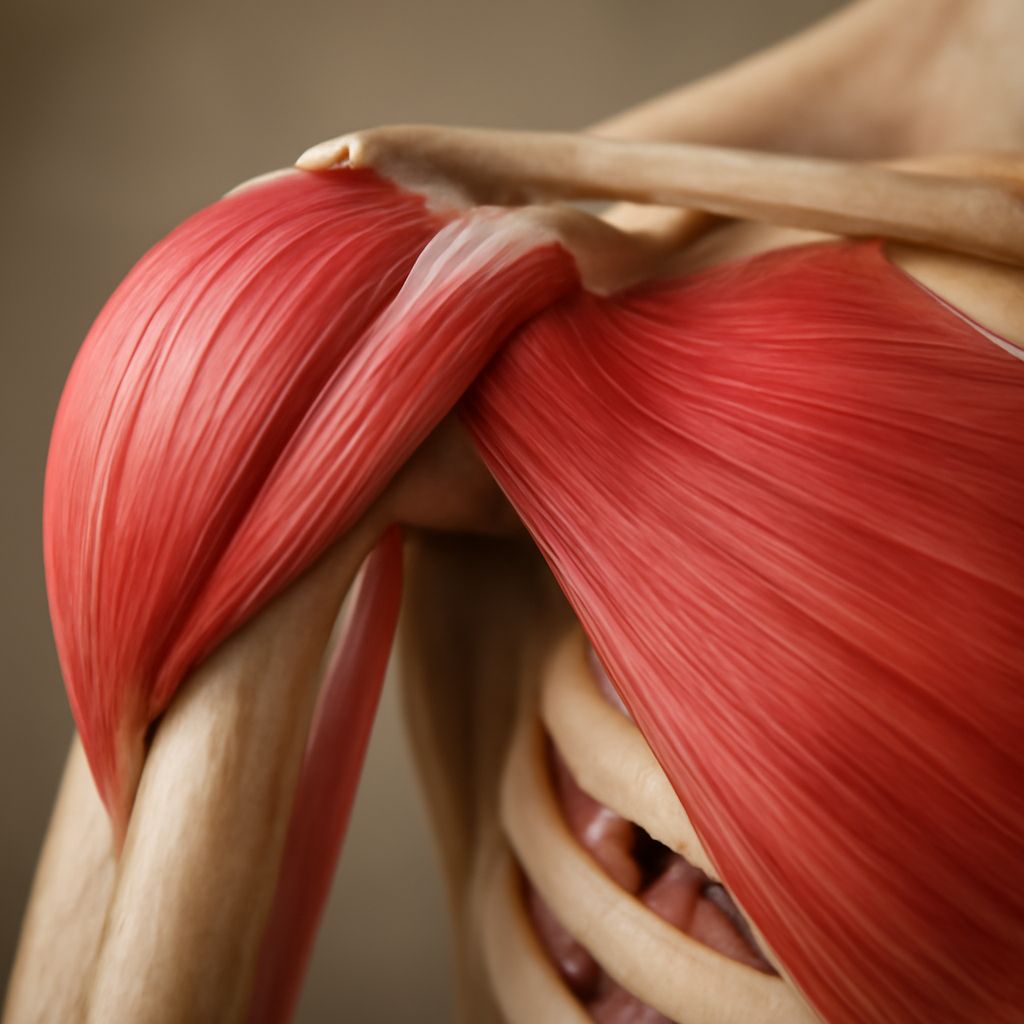
Deltoid and Surrounding Muscles
The deltoid covers the entire outer shoulder and is the primary mover for raising the arm to the side and front. Beyond the rotator cuff and deltoid, muscles like the trapezius, serratus anterior, and pectoralis minor all contribute to how the shoulder blade moves and how force transfers between your arm and trunk. These supporting muscles are often overlooked in rehabilitation but are essential to full function.
Nerves and Blood Supply
Brachial Plexus Overview
The brachial plexus is a network of nerves that originates from the cervical spine (C5 through T1) and branches out to supply the entire arm and shoulder. Damage to the brachial plexus, whether from trauma, compression, or inflammation, can cause weakness, numbness, or pain radiating down the arm. The axillary nerve, a branch of the brachial plexus, specifically controls deltoid function and sensation over the outer shoulder.
Blood reaches the shoulder primarily through the axillary artery, which runs through the armpit and gives off branches to supply the rotator cuff muscles, the joint capsule, and surrounding soft tissue. Adequate blood supply is especially important for tendon health, since tendons have limited circulation and heal slowly after injury.
Why Shoulder Anatomy Matters for Movement
Range of Motion
The shoulder achieves the greatest range of motion of any joint in the body, capable of moving through roughly 180 degrees of elevation and more than 60 degrees of internal and external rotation. This mobility depends on all the bones, joints, muscles, and nerves described above working in coordination. When one element is compromised, the rest must compensate, which often leads to secondary problems over time.
Common Injuries and Their Causes
Most shoulder injuries come from either acute trauma or repetitive overuse. Rotator cuff tears, labral injuries, and dislocations are among the most frequent problems seen in clinical practice. Understanding shoulder anatomy helps you recognize why certain activities carry specific risks and why certain exercises protect the joint better than others. For example, keeping the elbow below shoulder height during pressing movements reduces impingement risk by preserving space for the supraspinatus tendon.