Shoulder Anatomy Bones: Understanding the Skeletal Framework of the Shoulder
How does the shoulder manage to move in so many directions while still supporting the weight of the arm? The answer lies in the arrangement of the shoulder anatomy bones and how they connect to each other and to the rest of the skeleton. Unlike the hip joint, which sacrifices mobility for stability, the shoulder prioritizes range of motion, relying on muscles and ligaments rather than deep bony sockets to stay secure.
Understanding posterior shoulder anatomy, posterior leg anatomy for comparison, and even dog shoulder anatomy helps put the human shoulder in a broader biological context. Each of these skeletal arrangements reflects a different set of mechanical priorities. Posterior skull anatomy also connects to the shoulder through the cervical spine, making the shoulder part of a continuous chain of structures that runs from the back of the head to the fingertips.
Overview of Shoulder Anatomy Bones
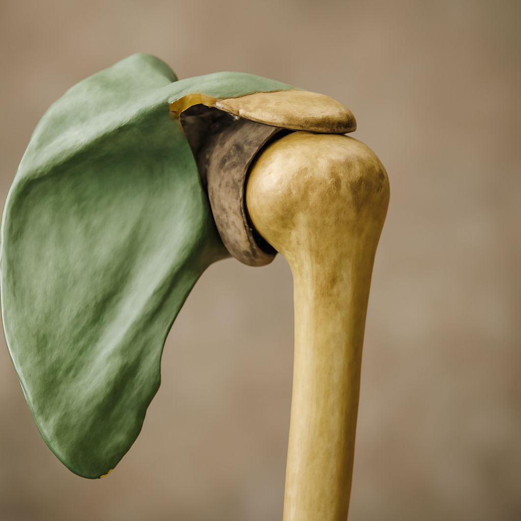
Three bones make up the shoulder complex: the clavicle (collarbone), the scapula (shoulder blade), and the humerus (upper arm bone). The clavicle runs horizontally from the sternum to the tip of the shoulder. The scapula is a flat, triangular bone sitting against the posterior rib cage. The humerus connects below, its rounded head fitting into the glenoid cavity of the scapula to form the primary shoulder joint. The shoulder anatomy bones work as a unit, and a change in position or condition of any one affects the others.
Posterior Shoulder Anatomy: The Back View
Viewing the shoulder from behind, the most prominent feature is the spine of the scapula, a bony ridge that runs diagonally across the posterior surface and terminates at the acromion process. The posterior shoulder anatomy also includes the infraspinous fossa below the spine of the scapula and the supraspinous fossa above it. Both fossae house rotator cuff muscles. The posterior glenohumeral joint capsule, though soft tissue rather than bone, limits excessive forward movement of the humeral head and is a common site of tightness in overhead athletes.
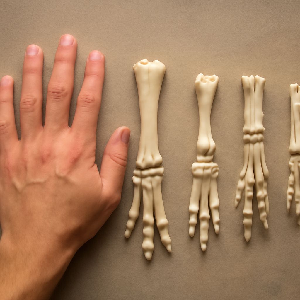
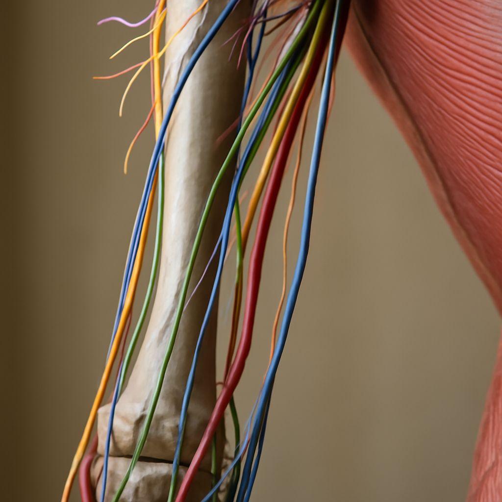
Comparing Posterior Leg Anatomy to the Shoulder
The posterior leg anatomy offers a useful comparison to the shoulder, specifically in how different ball-and-socket joints handle the same engineering problem. The hip socket (acetabulum) is much deeper than the glenoid, wrapping more than halfway around the femoral head. This depth provides inherent stability that the shoulder lacks. In the posterior leg anatomy, the hamstring muscles control hip extension much like the posterior rotator cuff controls shoulder external rotation. Both regions use a combination of ligamentous restraint and muscular co-contraction to protect the joint during dynamic movement.
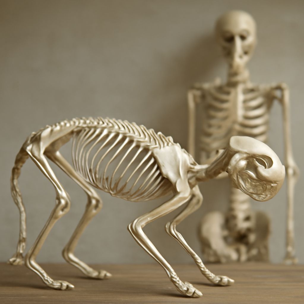
Dog Shoulder Anatomy: How It Differs from Humans
Dog shoulder anatomy lacks the clavicle entirely, or has only a vestigial remnant of one. This is not a deficiency; it is an adaptation for running. Without a clavicle connecting the shoulder to the sternum, the dog’s shoulder blade can move freely over the rib cage, allowing a longer stride length and more elastic energy storage during locomotion. The dog shoulder anatomy also places the scapula more parallel to the spine rather than perpendicular as in humans, which changes the mechanics of how the forelimb extends and retracts during a gallop. Veterinary professionals study dog shoulder anatomy extensively because shoulder injuries in working and sporting dogs carry significant welfare implications.
Posterior Skull Anatomy and Its Connection to the Shoulder
The posterior skull anatomy includes the occipital bone, which forms the back and base of the skull, and the mastoid processes of the temporal bones, which serve as attachment points for muscles that connect the head to the neck and shoulders. The trapezius muscle, one of the largest in the back, originates along the posterior skull anatomy from the superior nuchal line and the external occipital protuberance, then sweeps downward to attach to the clavicle, the acromion, and the spine of the scapula. This means that tension or restriction anywhere between the back of the skull and the shoulder blade directly affects shoulder movement.
Key Ligaments and Their Roles
Several ligaments stabilize the shoulder anatomy bones against each other. The coracoclavicular ligaments (conoid and trapezoid) hold the clavicle tightly to the coracoid process of the scapula. The glenohumeral ligaments (superior, middle, and inferior) reinforce the joint capsule at three specific points, each one resisting different directions of humeral displacement. The coracoacromial ligament forms a roof over the rotator cuff tendons. When this arch narrows due to posture or structural changes, the tendons underneath can become irritated, which is the mechanism behind impingement syndrome.
How the Bones Work Together in Movement
Shoulder movement is never isolated to a single joint. When you raise your arm overhead, the glenohumeral joint provides roughly 120 degrees of movement while the scapula rotates upward on the rib cage to contribute another 60 degrees. This scapulohumeral rhythm, as it is called in clinical anatomy, requires all three shoulder anatomy bones to move in precise coordination. A restriction in any joint, whether between the clavicle and sternum, the clavicle and acromion, or the humerus and glenoid, disrupts the whole pattern and typically produces pain at a location different from where the actual restriction sits.