Deer Skeleton Anatomy: Bones, Muscles, and Drawing Reference for Artists
Why do artists and naturalists study deer skeleton anatomy? Because understanding what sits beneath the surface tells you exactly why a deer moves and looks the way it does. A deer anatomy drawing that gets the bone placement wrong looks wrong in the finished piece, even if the viewer cannot articulate why. The proportional relationships between leg length, spine curve, and skull size are governed by skeletal structure, and learning that structure gives your drawings a believability that surface-level rendering alone cannot provide.
Beyond deer, this kind of study extends naturally into deer muscle anatomy for surface modeling, wolf muscle anatomy for comparative anatomy work, and even muscle anatomy games that make this technical content more accessible. Each of these areas connects to the same foundational practice: understanding how biological structures dictate visible form.
The Deer Skeleton: Key Structures and Proportions
Skull and Antler Attachment
The deer skull is elongated compared to many mammals, with a long facial region housing the teeth used for grazing. In male deer, antlers grow from permanent bony projections called pedicles on the frontal bone. When drawing deer skeleton anatomy from the front, the skull’s width relative to the width at the shoulder is a critical proportion to observe. Many anatomical deer drawings make the skull too large relative to the body, which reads as juvenile rather than adult.
Vertebral Column and Back Line
The deer’s spine shows a characteristic curve: relatively level through the thoracic region, then slightly rising toward the haunches. The withers, the high point at the base of the neck, are formed by the elongated spinous processes of the thoracic vertebrae. In a deer anatomy drawing, the withers define the highest point of the back and inform where the neck attaches to the body. Getting this landmark right organizes everything else in the body outline.
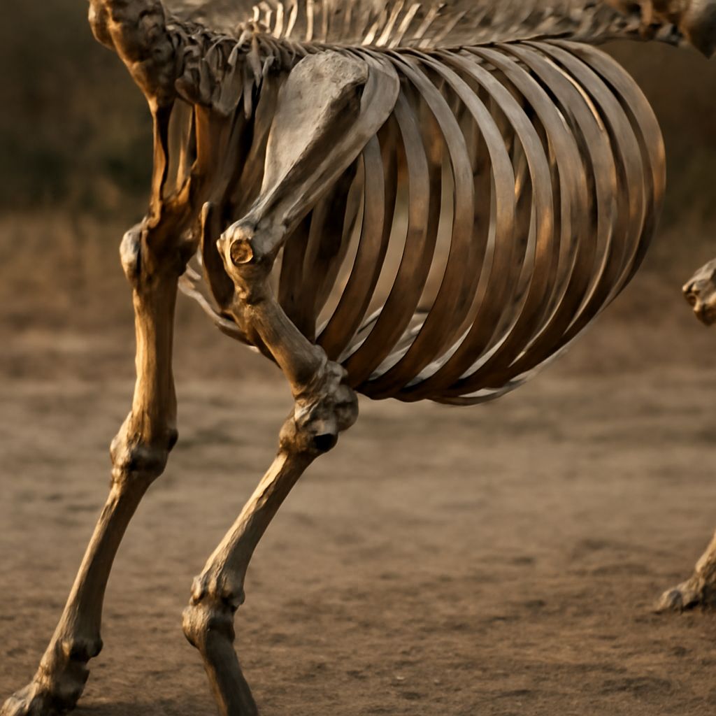
Limb Bones and Long Bone Proportions
Deer are digitigrade mammals, meaning they walk on the tips of their toes. What looks like a backward-bending knee in the rear leg is actually the heel, elevated high off the ground. The true knee in the rear leg is tucked close to the body and less visible in a standing pose. Understanding this limb structure is one of the most common corrections needed in deer skeleton anatomy for beginner artists.
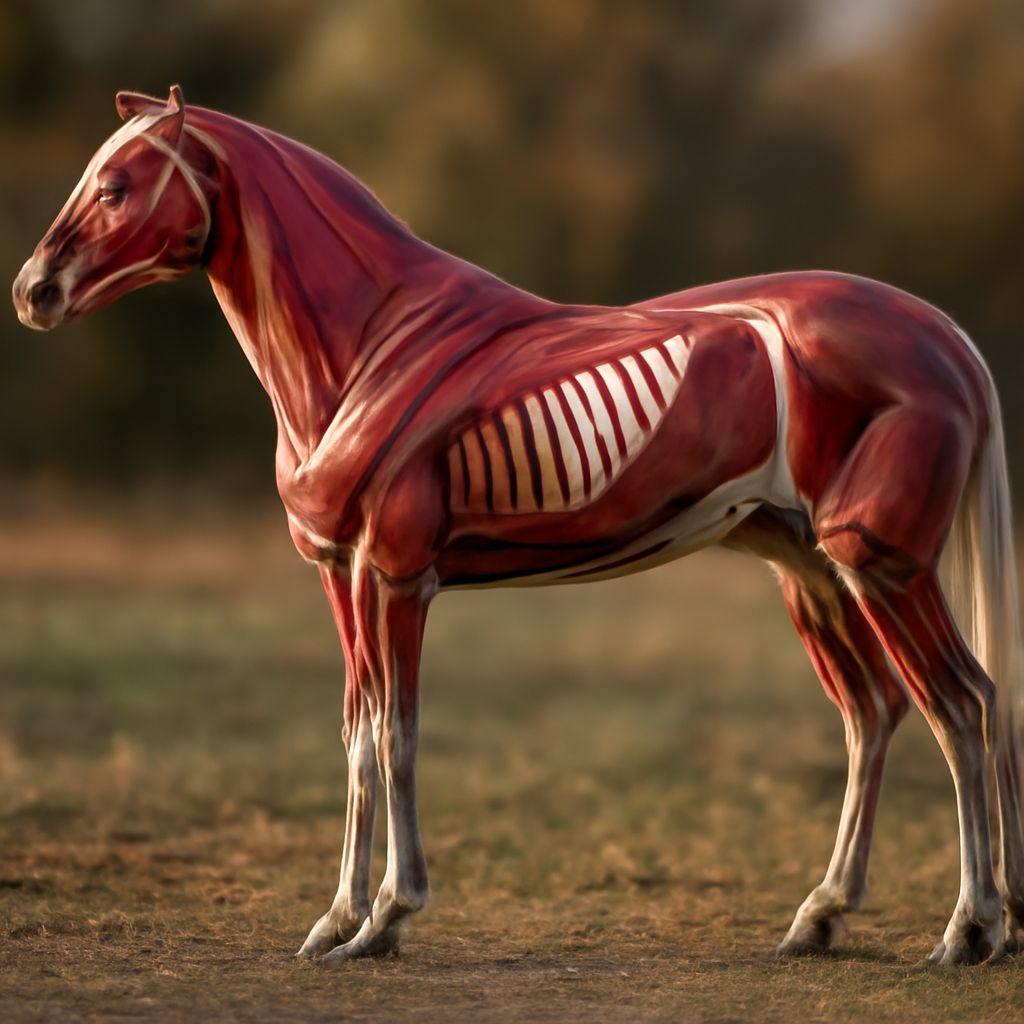
Deer Muscle Anatomy for Surface Form
Shoulder Muscles
Deer muscle anatomy at the shoulder shows the trapezius, deltoid, and triceps creating the distinctive triangular mass above the foreleg. This area in a living deer reads as a prominent muscle group that changes shape significantly as the leg moves forward and backward. When doing a deer anatomy drawing from life or reference, study the shoulder contour across multiple leg positions to understand how the surface changes.
Hindquarter Muscles
The hindquarters of a deer contain the most powerful muscle mass in the body, including the gluteus and hamstring groups that power jumping and sprinting. In deer muscle anatomy, these muscles create the rounded, full silhouette of the haunches visible in a three-quarter rear view. The muscle mass here is much greater than in the shoulder region, which creates the characteristic pear shape of the deer’s body when viewed from the side.
Neck Musculature
The neck muscles of a deer support a heavy skull and long antlers in males while allowing the head to reach low to the ground for grazing. The nuchal ligament, a strong elastic band running along the top of the neck, helps support the head’s weight. In deer anatomy drawing, the neck’s width should taper slightly from the head junction toward the chest rather than maintaining a uniform diameter, which reflects the underlying muscle distribution.
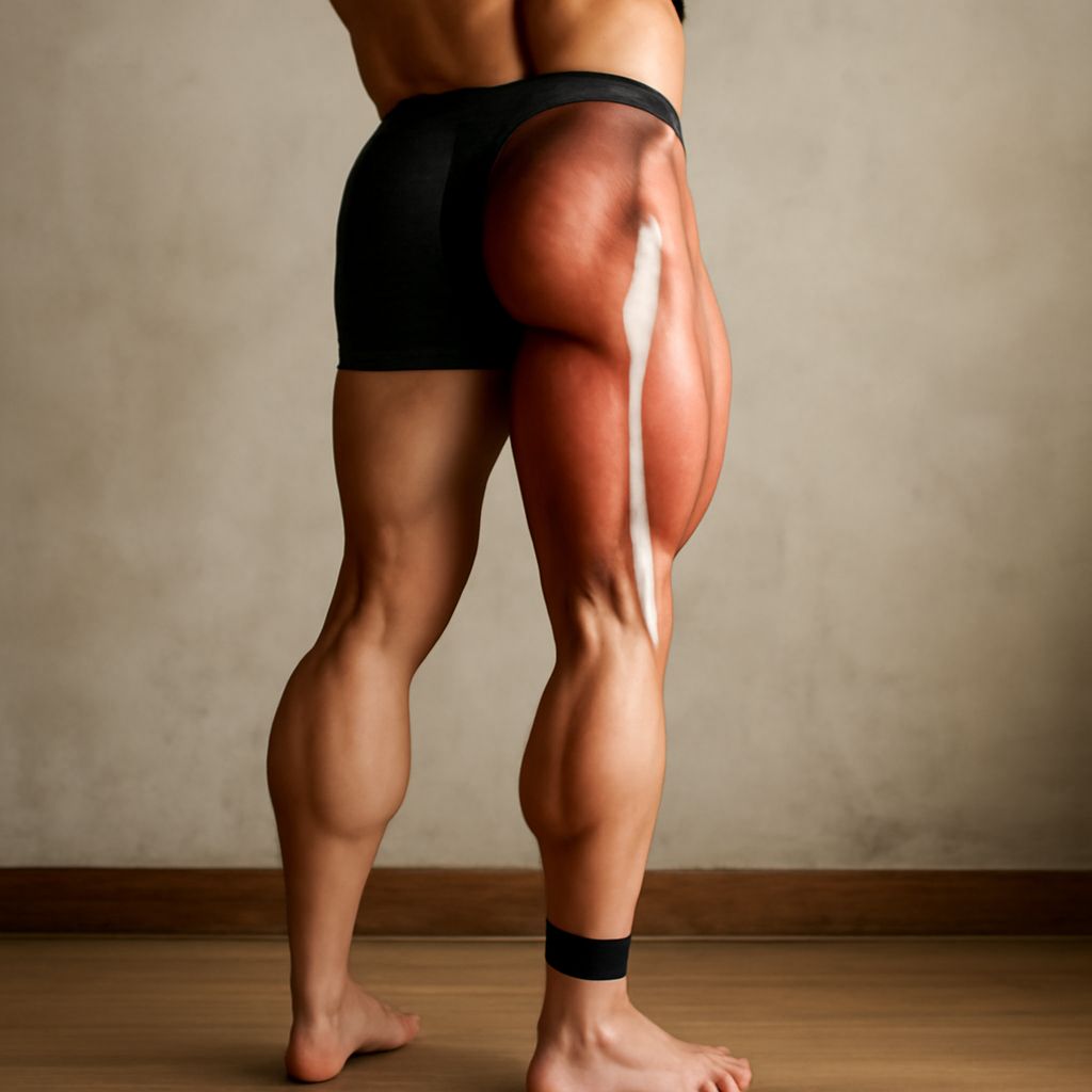
Wolf Muscle Anatomy: A Comparative Perspective
Key Differences from Deer
Wolf muscle anatomy reflects a different evolutionary strategy. Wolves are predators built for sustained pursuit over distance, while deer are prey animals built for explosive acceleration and jumping. Wolf muscle anatomy shows a deeper chest relative to the abdomen, longer limbs relative to body size, and more developed shoulder muscles for ground-covering trot mechanics. Comparing wolf and deer anatomy in a drawing context sharpens your ability to read species-specific body proportions rather than applying generic four-legged animal templates.
Surface Musculature Comparison
In wolf muscle anatomy, the latissimus dorsi creates a visible muscle mass behind the shoulder that reads as a distinct surface feature. In deer, this muscle is proportionally smaller and less prominently visible through the coat. Studying these differences in wolf muscle anatomy alongside deer anatomy drawing teaches you to see anatomical individuality in animals rather than applying one size fits all structural templates.
Muscle Anatomy Games for Interactive Learning
Digital Tools for Anatomy Study
Muscle anatomy games provide an interactive alternative to static diagrams for learning muscle placement and names. Several free web-based tools allow you to click on individual muscles in a three-dimensional model, see their names, and understand their attachment points. These tools work particularly well for deer muscle anatomy because they let you rotate the model to see how muscles appear from different viewing angles, which is essential reference for multi-perspective drawing.
Combining Games with Sketchbook Practice
The most effective approach combines muscle anatomy games with direct sketchbook study. Use the interactive tools to identify and name muscles, then draw simplified versions of those muscles from memory without the reference visible. Testing your recall through drawing consolidates the information more effectively than reviewing diagrams alone. Apply this method to both deer skeleton anatomy and wolf muscle anatomy to build a comparative reference library in your own sketchbooks.