Anatomical Drawings: From the Anatomical Heart Sketch to Leonardo Anatomy
What makes anatomical drawings different from other scientific illustrations? They sit at the intersection of precise observation and artistic skill, requiring the artist to render invisible internal structures in ways that communicate accurately to a viewer who cannot see the subject directly. An anatomical heart sketch is not just a picture of an organ; it is a teaching tool, a record of observation, and sometimes a work of art in its own right. The tradition of anatomical illustration stretches across centuries, from the detailed body illustration of medieval manuscripts to the revolutionary work of Leonardo anatomy that changed how educated Europeans understood the human form.
Anatomical illustration today serves medical education, scientific publishing, patient communication, and fine art equally well. The tools have changed from quill and metalpoint to digital brushes, but the core requirement remains identical: you must understand what you are drawing well enough to render it accurately, and you must render it clearly enough for someone else to learn from it. Whether you are creating an anatomical heart sketch, a full body illustration, or researching the influence of leonardo anatomy on contemporary medical art, that dual demand of knowledge and craft defines the field.
The History of Anatomical Drawings
Pre-Renaissance Medical Illustration
Early anatomical drawings relied on symbolic representation rather than direct observation. Organs were drawn according to inherited Greek and Arabic texts rather than from dissection. These illustrations communicated the location and function of organs as understood by the tradition, not as observed in actual specimens. The figures were schematic rather than realistic, which served the educational purpose of the era but limited the accuracy of the information conveyed.
Leonardo Anatomy and the Revolution of Observation
Leonardo da Vinci’s anatomical drawings, produced primarily between 1489 and 1513, changed the standard for what anatomical illustration could achieve. Leonardo anatomy combined direct observation from dozens of human dissections with extraordinary draftsmanship. His drawings of the heart, the fetus in utero, the muscles of the arm, and the structure of the skull remained unsurpassed in accuracy for generations. Leonardo anatomy introduced cross-sectional views, multiple angle representations of the same structure, and detailed notation that anticipated the conventions of modern technical illustration. His manuscripts were not published in his lifetime, which limited their immediate influence, but their rediscovery centuries later confirmed what contemporaries who knew the work already understood.
Vesalius and the Systematization of Anatomical Illustration
Andreas Vesalius published De Humani Corporis Fabrica in 1543, creating the first comprehensive atlas of human anatomy based on direct dissection. The body illustration in Vesalius was created by artists working in the studio of Titian, combining medical accuracy with Renaissance artistic quality. This combination of systematic science and artistic skill defined anatomical illustration for the next three centuries and established the standard that modern anatomical drawings still measure against.
The Anatomical Heart Sketch: Structure and Approach
Surface Anatomy of the Heart
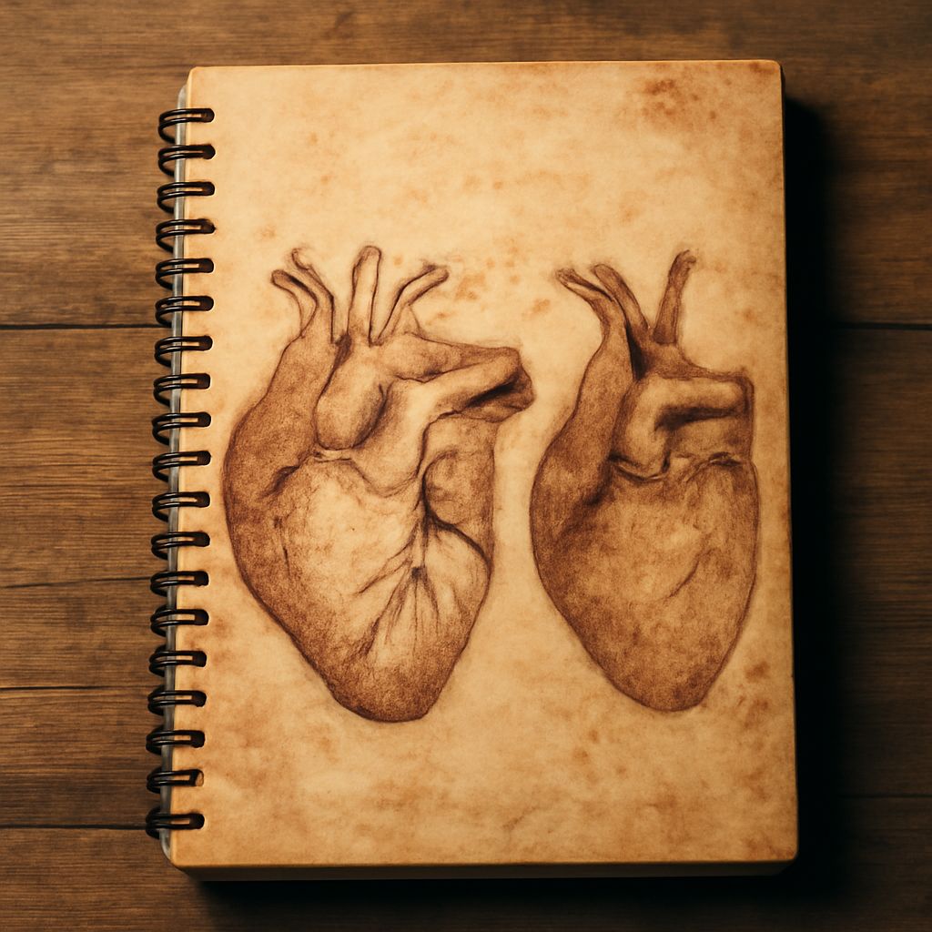
An anatomical heart sketch must accurately represent the heart’s external form before any internal structures are considered. The heart sits at roughly a 45-degree angle in the chest, with the apex pointing down and to the left. From the anterior view, the right ventricle forms most of the visible front surface, with the left ventricle making up the lower left portion and the two atria visible at the top. The great vessels, including the aorta, pulmonary artery, and superior vena cava, emerge from the superior aspect and define the top profile of the heart’s external form.
Internal Structure in Anatomical Illustration
When creating an anatomical illustration of the heart’s internal structure, the standard approach uses a coronal or sagittal section to reveal the chambers and valves simultaneously. The four chambers, two atria and two ventricles, need to be shown in their correct relative positions and proportions. The valves between chambers appear as flat structures at their respective openings. This kind of body illustration requires knowing the structure well enough to draw it correctly without being able to look at a cut heart directly.
Rendering Techniques for the Heart
The heart’s surface has a characteristic reddish-brown color in cadaveric specimens and a brighter red-pink in fresh tissue. For an anatomical heart sketch in graphite, tonal modeling using stippling or soft hatching creates the rounded muscular form. For a colored anatomical illustration, saturated reds and warm shadows suggest the cardiac muscle’s specific texture without being distractingly literal.
Body Illustration: Contemporary Approaches
Digital Tools in Anatomical Drawing
Contemporary anatomical drawings increasingly use digital tools, from 3D modeling software that generates accurate reference images from any angle to digital painting applications that simulate traditional media. These tools do not replace anatomical knowledge but they extend what is achievable in terms of rendering quality and the ability to produce multiple view variations quickly. A body illustration project that once required weeks of manual work can now be completed in days without sacrificing accuracy.
Combining Scientific Accuracy with Artistic Quality
The best contemporary anatomical illustration manages to be scientifically precise and visually compelling simultaneously. This balance is not easy to achieve and represents a genuine synthesis of two demanding disciplines. Artists who work in this field typically have either formal medical illustration training or deep independent study of anatomy combined with strong fine art foundations. Leonardo anatomy demonstrated that both are possible in a single practitioner; contemporary medical illustrators continue that tradition.
Learning Anatomical Drawing Today
Reference Resources
For anatomical drawings, the best references combine text and image in ways that explain not just what structures look like but how they relate to each other in three dimensions. Gray’s Anatomy, Netter’s Atlas, and the work of Eliot Goldfinger on animal anatomy all represent this standard. When creating an anatomical heart sketch or other anatomical illustration, always verify your drawing against multiple reference sources rather than relying on a single image.
Practical Study Sequence
Begin anatomical drawings study with the major external body forms before moving to internal structures. Understanding surface anatomy, what you can see and feel from outside the body, makes internal anatomical illustration more meaningful because you understand where the internal structures sit relative to the surface. Then progress from large-scale structures (major organ positions) to smaller-scale detail (vessel and nerve paths). This layered approach mirrors how anatomical illustration teaching has been structured since the Vesalian tradition.