Deer Anatomy: A Complete Guide to Bones, Organs, and Body Systems
What does a deer look like on the inside, and why does that matter? For hunters, wildlife managers, and artists, understanding deer anatomy changes how you interpret what you observe in the field and how you represent the animal in drawing or technical illustration. The anatomy of a deer is well-adapted for a prey animal that relies on speed, keen senses, and efficient digestion of plant material. Every major system, from the skeletal framework to the organ placement, reflects these evolutionary priorities.
This guide covers the anatomy of deer in enough detail to be genuinely useful whether you are studying for a wildlife biology exam, preparing to field-dress a deer, or building an accurate deer anatomy chart for reference. The deer anatomy organs section is particularly relevant for hunters who want to understand shot placement and field dressing logistics, while the skeletal and muscular sections serve artists and naturalists who need accurate structural reference.
Skeletal Structure of the Deer
Skull and Dentition
Deer anatomy at the skull shows an elongated facial region with a wide muzzle, large eye sockets positioned on the sides of the head, and a dental formula adapted for grazing. Deer lack upper incisors, instead having a hard dental pad that the lower incisors press against when cropping vegetation. This anatomical detail is visible in a deer anatomy chart that shows the skull in lateral view. The anatomy of a deer skull also includes the pedicles, bony projections of the frontal bone from which antlers grow annually in male deer.
Vertebral Column
The deer anatomy vertebral column has seven cervical vertebrae (the same number as in virtually all mammals, including humans), 13 thoracic vertebrae with corresponding ribs, six or seven lumbar vertebrae, four fused sacral vertebrae, and a variable number of caudal vertebrae in the tail. The thoracic vertebrae have elongated spinous processes at the withers region, creating the characteristic ridge at the base of the neck that is clearly visible in both living animals and deer anatomy charts used for art reference.
Limb Bones
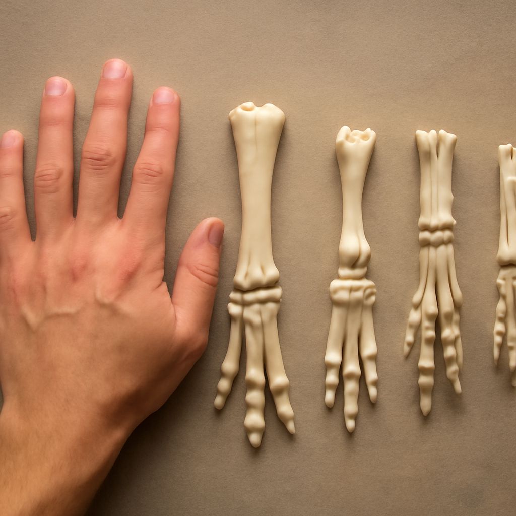
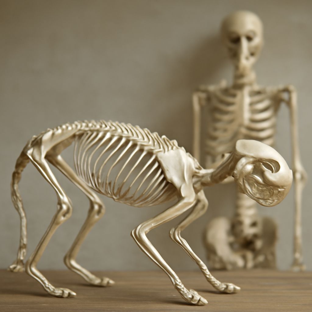
Deer are even-toed ungulates, walking on two functional toes (the third and fourth digits) on each foot, with vestigial dewclaws (the second and fifth digits) elevated above the ground. In the anatomy of a deer, the long bones of the leg include the femur, tibia, metatarsus, and the two main phalanges of each functional toe. The apparent “backward knee” in the rear leg is the hock joint, which corresponds to the human ankle and heel. The actual knee sits close to the body and is less visually prominent in a standing deer.
Deer Anatomy Organs: The Major Systems
Digestive System
Deer anatomy organs include a four-chambered ruminant stomach: the rumen, reticulum, omasum, and abomasum. This system allows deer to consume large quantities of vegetation quickly, then regurgitate and rechew (ruminate) it at leisure in a safe location. The rumen is the largest chamber and in a mature deer can hold 8 to 10 liters of partially digested material. Understanding deer anatomy organs in the abdominal region is essential for field dressing, since puncturing the rumen contaminates the meat.
Cardiovascular and Respiratory
The deer anatomy organs of the thoracic cavity include the heart, which sits in the pericardial sac between the lungs and just behind the sternum, and the two lungs, which fill the majority of the thoracic space. In a deer anatomy chart for hunting reference, the heart-lung zone is typically marked as the primary target for an ethical, effective shot. The heart sits lower in the chest than many hunters expect, roughly at the level of the fifth or sixth rib in a standing deer.
Liver and Kidneys
The liver sits in the anterior abdomen, just behind the diaphragm on the right side. In the anatomy of a deer, the liver is one of the largest organs and shows clearly in field dressing because of its dark red-brown color and its position immediately adjacent to the diaphragm. The kidneys sit in the dorsal abdominal region on either side of the spine, embedded in perirenal fat. Understanding the anatomy of deer organ placement helps with field dressing efficiency and with understanding post-shot behavior in hunting contexts.
Muscular System for Artists
Major Surface Muscles
The deer anatomy chart most useful for artists focuses on the surface muscles that define the animal’s visible contours. The trapezius creates the neck-to-shoulder slope. The gluteus and hamstring group defines the haunches. The triceps creates the back of the foreleg. The deltoid and infraspinatus create the shoulder mass. These six muscle groups, clearly understood, account for most of what you see when you look at a deer from the side and three-quarter views that are most common in wildlife illustration.
How Muscles Change with Movement
The anatomy of deer changes visibly as the animal moves. The shoulder muscles shift forward as the foreleg extends, creating a more prominent mass in front of the leg. The hindquarter muscles bunch and contract as the rear leg drives backward during a gallop. Understanding these dynamic changes in deer anatomy allows you to draw the animal in motion convincingly, which static reference photos alone do not fully prepare you for.