Tooth Anatomy: A Complete Guide to Dental Structure and Function
Do you know what’s actually inside a tooth? Most people have a rough sense that teeth have roots and some kind of inner layer, but the full picture of tooth anatomy is more intricate — and more relevant to your daily dental decisions — than that. Understanding dental anatomy helps you make sense of why certain symptoms appear, what happens during specific procedures, and how different parts of a tooth respond to damage or decay. That knowledge gives you a much more productive conversation with your dentist.
This article walks through the complete structure of a tooth from crown to root tip. You’ll learn how dental anatomy connects to common clinical concerns, why molar anatomy differs from front-tooth anatomy, how dental infographics typically simplify these structures for patient education, and what the often-misunderstood term tooth fairy font has to do with the broader world of dental-themed design. By the end, you’ll have a clear, accurate picture of what each layer of your tooth actually does.
The Major Layers of a Tooth
Enamel
Enamel is the outermost layer of the crown — the visible portion of the tooth above the gumline. It’s the hardest substance the human body produces, composed almost entirely of hydroxyapatite, a crystalline calcium phosphate mineral. Despite its hardness, enamel is brittle and doesn’t regenerate once lost. Acid exposure from food, beverages, and bacterial byproducts gradually dissolves the mineral structure, which is why consistent oral hygiene and dietary choices have long-term consequences for enamel integrity.
Dentin
Beneath the enamel sits dentin, a yellowish tissue that makes up the bulk of the tooth’s volume. Dentin is less hard than enamel and contains microscopic tubules — tiny channels that run from the pulp outward. These tubules are why dentinal exposure causes sensitivity: temperature changes, sweet foods, or even air movement can stimulate the nerve endings inside the pulp through those channels. Understanding this aspect of tooth anatomy explains why sensitivity often increases after enamel wears away or gums recede.
Pulp
The pulp chamber sits at the center of the tooth, housing blood vessels, lymphatic tissue, and the nerve supply. This is the living core of the tooth. During tooth development, the pulp plays an active role in dentin formation. In a mature tooth, its primary function shifts to sensory detection — it registers temperature, pressure, and pain. When pulp tissue becomes infected or irreversibly inflamed, a root canal procedure removes it to preserve the remaining tooth structure.
Cementum and the Periodontal Ligament
Cementum covers the root surface and anchors the periodontal ligament — a network of collagen fibers that connects the tooth root to the surrounding alveolar bone. This ligament isn’t rigid; it acts as a shock absorber during biting and chewing. Understanding dental anatomy at this level explains why teeth feel sensitive after heavy grinding, and why orthodontic movement is possible: controlled pressure gradually reshapes the bone on one side of the ligament while new bone fills in behind.
Molar Anatomy vs. Front Tooth Anatomy
Not all teeth share the same internal architecture. Molar anatomy is particularly distinct because molars have multiple roots — typically two in lower molars and three in upper molars — each containing its own root canal. This multi-root structure is one reason molar root canals are more involved than those on single-rooted front teeth. The pulp chamber in a molar also has multiple canals branching from it, all of which must be cleaned and sealed during endodontic treatment.
Front teeth (incisors and canines) have a single, usually straight root and a simpler pulp chamber. Their crown shape is optimized for cutting and tearing rather than grinding, which is why the biting edge is thin and blade-like rather than cusped. When examining molar anatomy versus incisor anatomy, the functional differences in crown shape map directly to differences in root number, root length, and the complexity of the internal canal system.
How Dental Infographics Present Tooth Structure
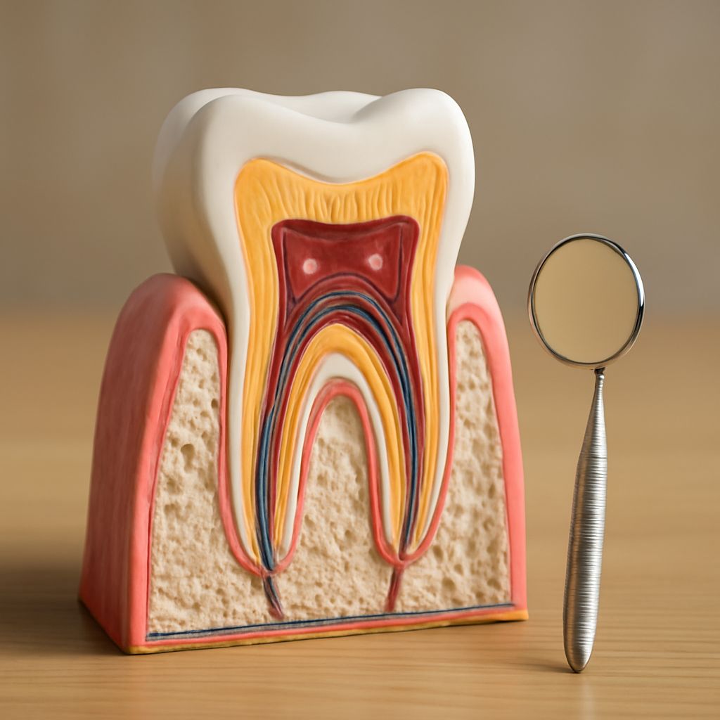
Dental infographics are a staple of patient education materials in clinics, schools, and public health campaigns. A well-designed infographic on tooth anatomy typically shows a cross-section view with labeled layers, often using color coding to distinguish enamel, dentin, pulp, cementum, and bone. The cross-section format works well because it reveals what’s otherwise invisible to patients — most people only see the crown surface and have no mental model for what happens below the gumline.
The challenge with dental infographics is balancing accuracy against readability. A fully accurate anatomical diagram includes details that would overwhelm a general audience — multiple canal variations in molar anatomy, accessory canals, the distinction between coronal and radicular pulp. Effective infographics simplify these details without introducing misconceptions, using callout lines and brief labels rather than dense paragraphs.
Tooth Fairy Font and Dental Design
If you’ve searched for resources in dental-themed design, you’ve likely encountered the term tooth fairy font — a category of playful, child-friendly typefaces used in pediatric dentistry materials, educational worksheets, and character-based educational media. These fonts typically feature rounded letterforms, soft serifs, and sometimes incorporate tooth or smile shapes into the letterforms themselves. They’re a deliberate contrast to the clinical typography used in adult dental materials.
The choice of a tooth fairy font isn’t just aesthetic. Research in pediatric healthcare communication suggests that visual design elements — including typeface — affect how anxious young patients perceive a clinical environment. Warm, approachable typography in waiting room signage and educational materials can reduce procedural anxiety, which makes the clinical experience easier for both child and clinician. That’s a practical argument for taking typeface choices in dental design seriously.
Putting It Together: Why Tooth Anatomy Knowledge Matters
A working understanding of tooth anatomy changes how you experience dental care. When a dentist mentions dentin exposure, you understand why the sensitivity is happening. When a treatment plan includes a crown to protect a cracked tooth, you know what structure is being preserved. When molar anatomy comes up in a discussion about an extraction, you have a framework for understanding why that specific tooth presents a different clinical situation than a front tooth.
Dental anatomy knowledge also helps you evaluate the dental infographics and educational materials you encounter. Accurate patient education resources will show layered anatomy in cross-section, label cementum and the periodontal ligament, and represent molar root complexity honestly. Materials that skip these details may be oversimplified to the point of being misleading.