Horse Anatomy: A Complete Visual Guide for Artists and Equestrians
What makes a horse drawing look alive, or a sculpture feel structurally correct? Understanding horse anatomy is the answer — whether you’re an equestrian assessing your animal’s health or an artist trying to capture movement on paper. The skeletal and muscular architecture of the horse is both elegant and complex, and a good horse anatomy diagram can reveal relationships between bones and muscles that aren’t obvious from observation alone. Knowing the anatomy of a horse changes how you see the animal entirely.
This guide covers the essential systems you need to understand, from skeletal structure to muscle groups. You’ll work through anatomy of horse terminology, regional landmarks, and how each region functions during movement. The anatomy of the horse rewards careful study, and the knowledge you gain transfers directly to better horsemanship and more accurate artistic representation.
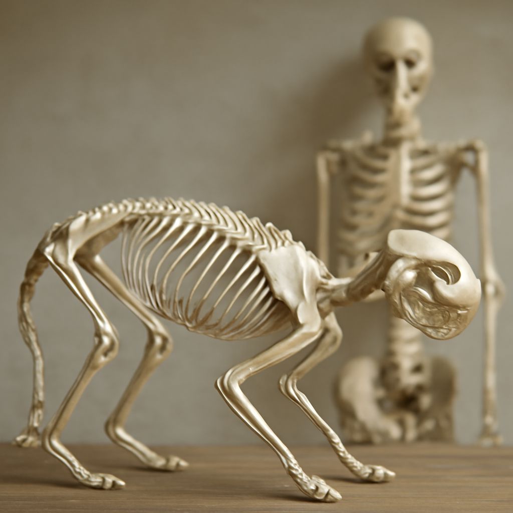
The Skeletal Framework
A horse’s skeleton contains roughly 205 bones, depending on variations in the number of lumbar vertebrae and the presence or absence of certain small bones in the foot. The skeleton provides the framework for everything else — muscle attachment points, organ protection, and the levers that drive locomotion.
The Skull and Cervical Vertebrae
The horse skull is elongated to accommodate the large teeth and the wide, laterally placed eyes that give horses their panoramic vision. The seven cervical vertebrae of the neck are broad and allow the head remarkable range of motion. Understanding this region helps riders assess neck flexibility and helps artists draw the graceful arc of the equine neck accurately.
The Thoracic and Lumbar Spine
The thoracic vertebrae — eighteen in most horses — each bear a pair of ribs that form the rib cage. The lumbar vertebrae, typically six, connect the thorax to the pelvis. This lumbar-thoracic junction is the area where back pain most often develops, and any horse anatomy diagram worth studying will highlight this region clearly.
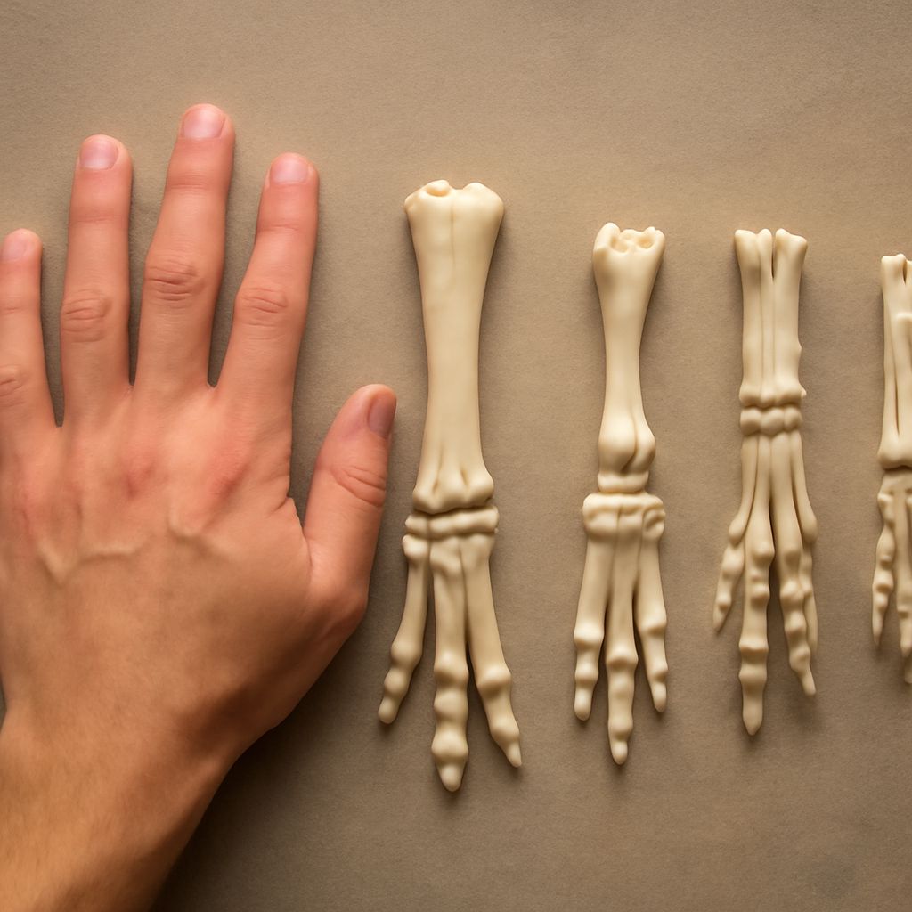
The Limbs and Feet
Each limb ends in a single toe — the hoof — making the horse a highly specialized ungulate. The forelimb lacks a true collarbone, relying instead on musculature to attach the shoulder to the trunk. The hindlimb drives propulsion. The hock joint in the hindleg corresponds to the human ankle, a fact that surprises many people seeing a detailed anatomy of a horse comparison diagram for the first time.
Major Muscle Groups
Muscle covers the skeleton in distinct groups that correspond to specific movements. For artists, understanding where muscles originate and insert explains why the horse’s silhouette changes so dramatically between standing, trotting, and galloping.
The Shoulder and Forearm Muscles
The trapezius, rhomboid, and serratus ventralis muscles attach the shoulder blade to the spine and rib cage. The triceps and biceps govern the elbow. When you see a horse reaching forward in a trot, you’re watching these muscles working in coordinated sequence. A quality study of horse anatomy will include cross-section views showing layered muscle depth.
The Hindquarter Power System
The gluteal muscles, hamstrings, and quadriceps of the hindquarter generate the thrust that propels the horse forward. The gluteus medius is the largest muscle in the horse’s body and defines the rounded, powerful shape of the croup. Weakness here directly affects collection and impulsion, key concepts in dressage. Any complete reference on anatomy of horse musculature will dedicate significant space to this region.
The Back and Abdominal Core
The longissimus dorsi runs along either side of the spine from neck to hindquarters and is the primary back muscle. Its condition correlates with the horse’s ability to carry a rider correctly. The abdominal muscles support the weight of the intestines and contribute to the rounding of the back during collection. These are regions where conditioning work makes a visible and measurable difference.
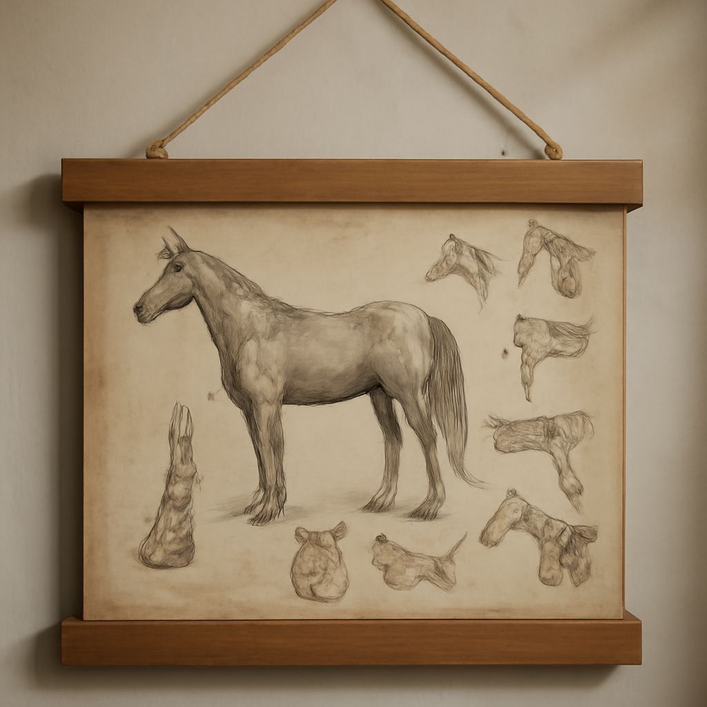
External Landmarks and Surface Anatomy
Surface anatomy — what you can see and feel from the outside — bridges the gap between internal structure and practical observation. The withers, the point of the hip, the point of the buttock, and the chestnut are all external landmarks that correspond to underlying skeletal or soft tissue structures.
When assessing a horse’s conformation, you evaluate how these landmarks relate to each other. A horse with a long, sloping shoulder will have different movement characteristics than one with an upright shoulder. Artists rendering the anatomy of the horse should memorize at least eight to ten external landmarks before attempting to draw the animal from life, as these points anchor proportion correctly.
Pro Tips Recap
Study both skeletal and muscular layers of horse anatomy — one without the other gives an incomplete picture. Use a layered horse anatomy diagram that lets you toggle between systems. If you’re an artist, practice drawing the skeleton first, then add muscle groups, then the skin surface. This method, used in classical anatomy study, produces far more accurate results than copying photographs directly. For equestrians, pairing visual anatomy study with hands-on palpation of your own horse makes landmarks concrete and unforgettable.