Anatomy Lab Essentials: From Moth Anatomy to Cactus Structure
What do a moth, a dog, a cactus, and the human voice have in common? Each becomes dramatically more understandable when you break it down systematically. An anatomy lab approach — whether in a formal educational setting or through self-directed study — transforms vague familiarity into specific, applicable knowledge. Moth anatomy reveals how insects achieve flight and camouflage with bodies far simpler than vertebrate equivalents. Canine muscle anatomy helps trainers, veterinarians, and artists understand how dogs generate power and express body language. The structures behind speech anatomy explain how humans produce the sounds of language. Even cactus anatomy tells a story of extreme adaptation to scarcity.
This guide takes an anatomy lab perspective across all four systems, providing the vocabulary and structural understanding that makes each topic more accessible for students, artists, and curious learners alike.
Moth Anatomy: Structure for Flight and Survival
Moths belong to the order Lepidoptera alongside butterflies. Their body plan follows the standard insect layout — head, thorax, abdomen — but the details of each region reveal specialized adaptations for nocturnal life.
The Head and Sensory Systems
A moth’s head carries compound eyes that detect movement and light levels, but its most remarkable sensory organs are the antennae. Unlike butterfly antennae, which are thin with clubbed tips, moth antennae are often feathered or comb-like — maximizing surface area for detecting pheromone molecules at extremely low concentrations. Moth anatomy here shows how structure follows function: the broader the antenna, the more receptors available.
Wings and Flight Mechanics
Moths have four wings — two forewings and two hindwings — connected during flight by a hook-and-loop structure called the frenulum. Wing scales, which give moth anatomy its characteristic dusty texture, are modified hairs that carry the pigment patterns used for camouflage or warning coloration. The wing venation pattern — the network of hollow tubes that stiffen each wing — is a key identification feature in moth taxonomy.
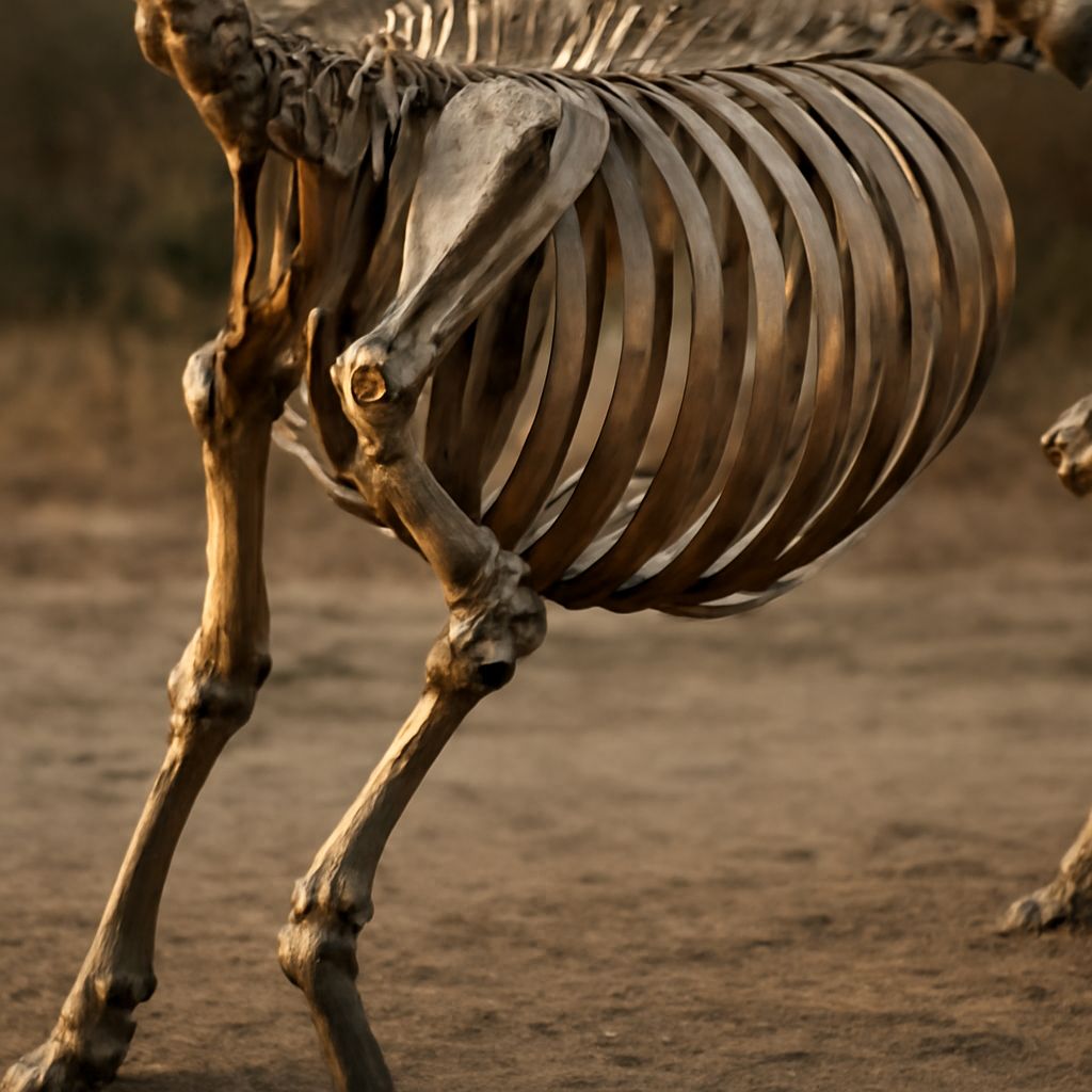
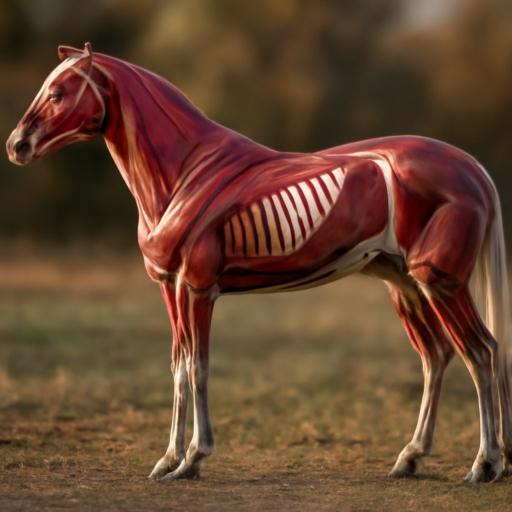
Canine Muscle Anatomy for Trainers and Artists
Dogs have over 700 muscles, but the functionally and visually significant ones number far fewer. Understanding canine muscle anatomy helps trainers assess fitness, identify injury patterns, and design appropriate conditioning programs. For artists, the same knowledge produces accurate, expressive animal drawings.
The trapezius and rhomboid muscles of the shoulder govern how a dog’s front legs swing forward and bear weight. The biceps femoris and semitendinosus in the hindquarter drive propulsion. The deep paraspinal muscles running along the spine maintain posture and contribute to core stability. Canine muscle anatomy follows the same functional logic as human anatomy — muscles cross joints to create movement — but the proportion and arrangement reflect four-legged locomotion rather than bipedal movement.
Speech Anatomy: The Voice Production System
Speech anatomy involves several distinct structures working in coordinated sequence. The respiratory system provides the airstream; the larynx produces initial sound via vocal fold vibration; the pharynx, oral cavity, and nasal cavity shape that sound into recognizable speech sounds; and the articulators — tongue, lips, teeth, and palate — fine-tune the sound into specific consonants and vowels.
The vocal folds in the larynx are perhaps the most mechanically sophisticated part of speech anatomy. They open and close approximately 100 to 300 times per second during voiced speech, and their tension determines pitch. Understanding this system helps singers, actors, speech therapists, and teachers work more effectively with voice production. An anatomy lab examination of laryngeal structure — even through models or diagrams — makes the abstract mechanics of voice immediately concrete.
Cactus Anatomy: Adaptation to Extremes
Cactus anatomy is a study in how evolution remakes standard plant structures for completely different functions. What appear to be leaves on a cactus are actually modified leaves called glochids or spines — they’ve lost their primary photosynthesis role and now function primarily for water conservation and defense.
The green, fleshy body of most cacti is actually the stem, which has taken over the photosynthesis function that leaves perform in other plants. This stem stores water in specialized parenchyma tissue, giving cacti their characteristic thick, succulent texture. The root system of cactus anatomy is typically shallow and wide-spreading, designed to capture rainfall quickly before it evaporates. In an anatomy lab context, cross-sections of cactus tissue reveal the distinct outer epidermis, the chlorophyll-containing cortex, and the central water storage core in a way that makes these adaptations visible and memorable.
The areoles — small, cushion-like structures unique to cacti — are modified short shoots from which spines, flowers, and new stem segments grow. Every spine, every flower, and every branching point on a cactus emerges from an areole. Recognizing areoles helps you correctly identify cactus anatomy features that might otherwise seem random or confusing.