Horse Leg Anatomy: Front Leg, Nerve Anatomy, Insect Leg, and Reference Drawing
Why does the horse leg anatomy topic attract such a wide range of searchers — equestrians, veterinarians, artists, and biology students? Because the horse leg is anatomically remarkable in ways that connect to broader questions about vertebrate evolution, locomotion efficiency, and animal structure. Horse leg anatomy reveals one of evolution’s most elegant solutions to the locomotion demands of a large, fast, cursorial animal. Horse front leg anatomy is particularly educational because the forelimb’s lack of a collarbone creates a unique suspension system that most people find surprising. Leg nerve anatomy in horses has direct clinical relevance for lameness diagnosis. The comparison with insect leg anatomy illuminates how fundamentally different solutions have evolved for the same basic locomotion problem across very different animal groups. And a quality leg reference drawing combines observational accuracy with structural understanding in ways that serve artists across multiple subject areas.
This guide covers each of these dimensions of leg anatomy with the depth that makes the knowledge practically useful.
Horse Leg Anatomy Overview
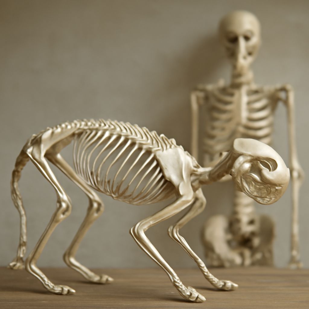
Horse leg anatomy begins with the basic recognition that what appears to be the horse’s knee in the front leg is anatomically the wrist — the carpus — while what appears to be the knee in the rear leg is anatomically the stifle — the equivalent of the human knee. This anatomical reorientation changes how you understand every structure in the horse’s limb and is often the first surprising insight students encounter when beginning equine anatomy study.
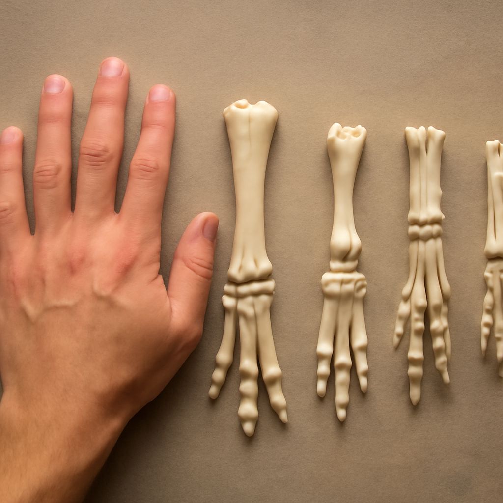
The horse’s single toe (the third digit), enclosed in the hoof, is the terminal element of a limb that has reduced all non-essential bones to stubs or vestigial structures — the splint bones, remnants of the second and fourth metacarpals — over evolutionary time. This extreme specialization for forward-backward locomotion in a single plane makes the horse’s limb one of the most efficient running systems in the mammalian kingdom.
Horse Front Leg Anatomy
Horse front leg anatomy is unusual among quadrupeds because the forelimb lacks a direct bony connection to the axial skeleton — there is no clavicle (collarbone) connecting the shoulder blade to the sternum as in humans. Instead, the entire front end of the horse is suspended in a muscular sling formed by the serratus ventralis, trapezius, and other muscles. This muscular suspension system acts as a spring, absorbing the impact forces of the forelimbs during locomotion and storing energy for release in the next stride.
The major bones of horse front leg anatomy from top to bottom: the scapula (shoulder blade), humerus (upper arm), radius and ulna (fused in the adult horse to form the forearm), the carpal bones (the equivalent of wrist bones), the large cannon bone (third metacarpal), the long and short pastern bones (first and second phalanges), and the coffin bone (third phalanx) within the hoof capsule. Each joint in this sequence has specific flexibility ranges that define the limb’s movement pattern.
Leg Nerve Anatomy for Lameness Diagnosis
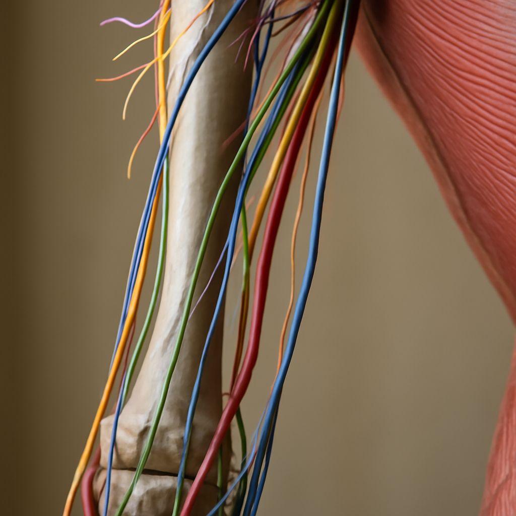
Leg nerve anatomy in horses is clinically significant because nerve blocks — the injection of local anesthetic around specific nerves — are the primary diagnostic tool for localizing the source of lameness. By systematically desensitizing regions of the foot and lower leg, veterinarians identify which specific structure, when its nerve supply is blocked, eliminates the lameness. This requires precise knowledge of exactly which anatomical region each nerve block desensitizes.
The major nerves in horse leg nerve anatomy: the palmar digital nerves run down either side of the pastern and desensitize the back of the foot when blocked. The palmar nerves desensitize a larger region of the lower leg. Sequential nerve blocks working up the limb allow triangulation of the pain source with great precision. Understanding this leg nerve anatomy system explains both the diagnostic process and why horses can develop specific lameness patterns that respond to specific blocking patterns.
Insect Leg Anatomy for Comparative Study
Insect leg anatomy provides a dramatically different evolutionary solution to the locomotion problem. Where the horse has evolved toward maximum limb reduction for straight-ahead speed, insects have retained six legs with multiple segments each — the coxa, trochanter, femur, tibia, and tarsus — that provide stability, traction, climbing ability, and versatile manipulation. The insect leg anatomy reflects a completely different set of ecological demands: most insects need to navigate complex three-dimensional surfaces, carry loads relative to their body weight, and perform multiple functional tasks with their legs beyond pure locomotion.
The comparison between horse and insect leg nerve anatomy and mechanical design illustrates a fundamental principle of comparative anatomy: homologous structures (corresponding structures from the same evolutionary origin) can be modified beyond recognition to serve completely different functions while retaining traceable structural relationships. Both the horse cannon bone and the insect femur are derivatives of the ancestral tetrapod femur, modified by hundreds of millions of years of different selective pressures.
Leg Reference Drawing for Artists
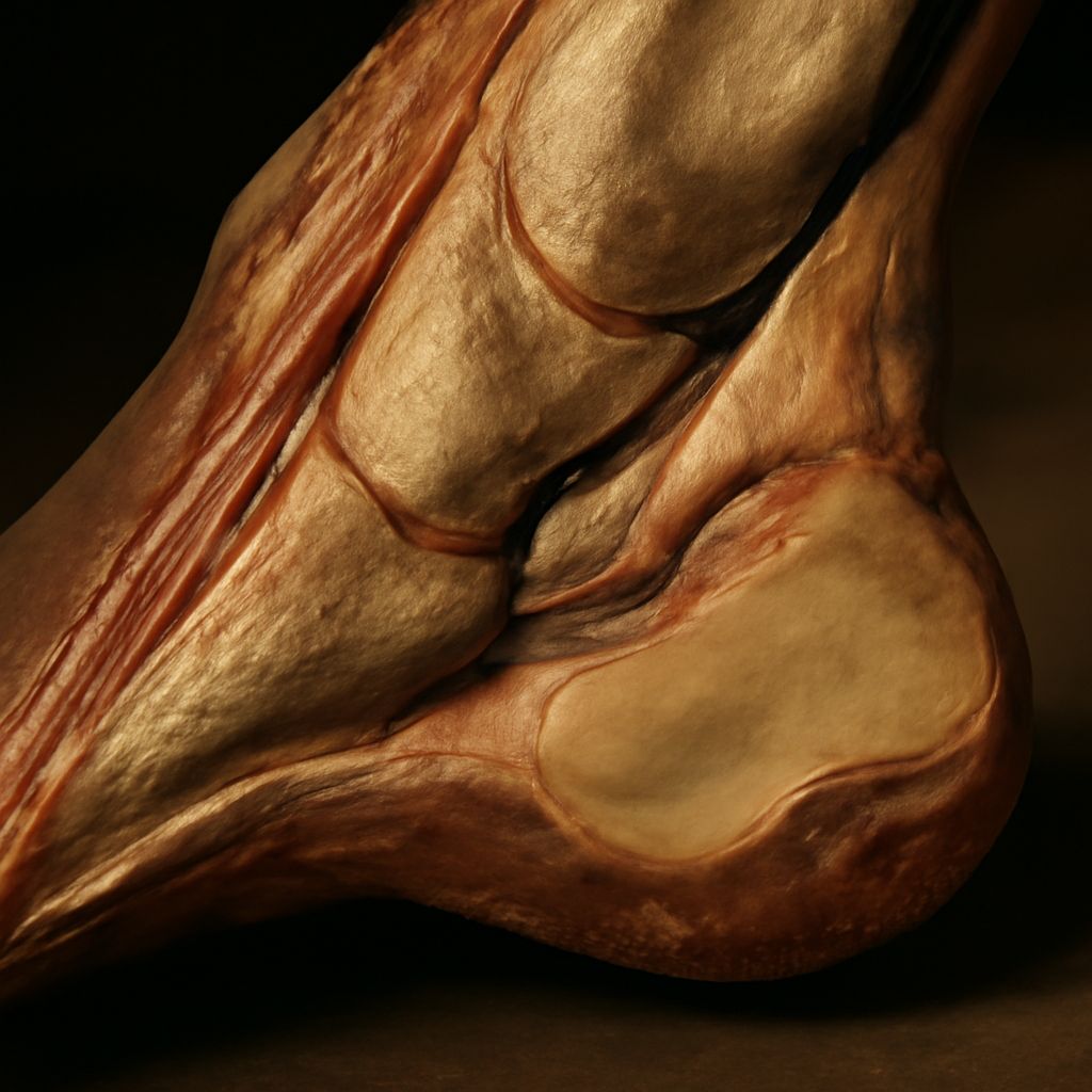
A quality leg reference drawing for artistic purposes captures both the surface appearance of the limb and enough structural information to make the drawing generative rather than just descriptive. The best leg reference drawing for equine artists shows multiple views — lateral, anterior, posterior, and medial — at the same scale, clearly labeled with major bony landmarks and muscle masses that create the visible surface form. These landmarks — the point of the elbow, the chestnut (horny growth on the inner leg), the ergot (horny growth at the back of the fetlock), and the heel bulbs — serve as spatial reference points that anchor proportion in complex or foreshortened views.