Female Dog Anatomy: A Complete Illustrated Guide
How well do you actually understand what’s happening inside your female dog’s body? Knowing female dog anatomy goes beyond basic biology — it helps you recognize health changes, communicate clearly with your vet, and make informed decisions about spaying or breeding. A good dog illustration can clarify structures that a written description alone cannot convey, which is why visual references play such an important role in veterinary education and owner awareness alike.
This guide walks you through the key body systems unique to female canines. You’ll learn about the female dog reproductive anatomy in plain language, understand why dog neck anatomy matters for handling and health assessment, and get a brief comparison with chicken reproductive anatomy to highlight how differently reproduction works across species. Real understanding starts with looking closely at what each structure actually does.
Key Body Regions and External Landmarks
Reading a Dog’s Body from the Outside
Before diving into internal structures, it helps to understand the external landmarks that guide physical examination. A dog illustration typically maps these landmarks clearly: the withers (top of the shoulder blades), the loin region behind the last rib, the flank, and the perineal area near the tail. These reference points matter because your vet palpates these zones during routine checkups to assess organ position and detect abnormalities.
In female dogs, the vulva is located below the anus in the perineal region. Its size and appearance change noticeably during estrus (heat), which is one of the earliest observable signs that a dog is entering her reproductive cycle. Knowing where to look — and what normal looks like — makes abnormalities easier to catch early.
Dog Neck Anatomy and Its Clinical Relevance
The neck contains the trachea, esophagus, jugular veins, carotid arteries, and the thyroid and parathyroid glands. Dog neck anatomy is relevant for a few practical reasons: the jugular vein is a common blood draw site, and the lymph nodes along the neck are among the first to enlarge when infection or illness is present.
For female dogs specifically, thyroid function can affect reproductive health — hypothyroidism has been linked to irregular estrous cycles and reproductive failure in some cases. Understanding the connections between neck anatomy and systemic health gives you a more complete picture of how your dog’s body functions as a whole.
Female Dog Reproductive Anatomy Explained
The Ovaries and Uterus
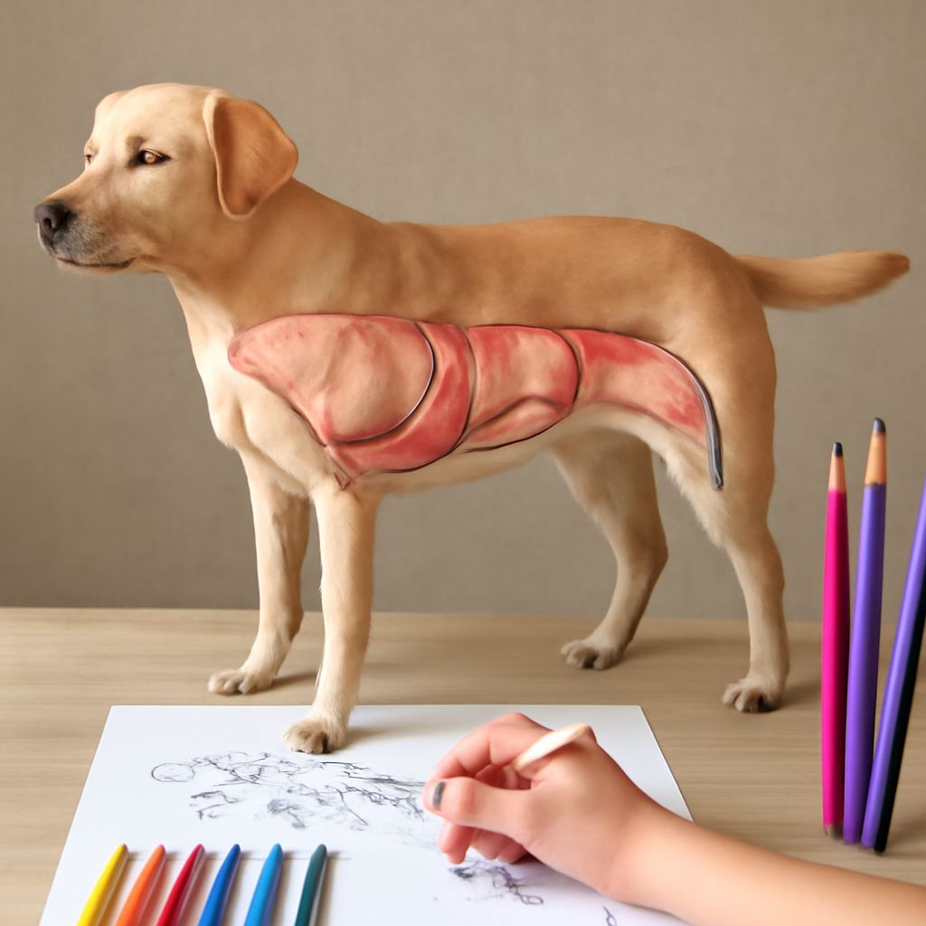
The female dog reproductive anatomy includes paired ovaries located just behind the kidneys. Each ovary is small — roughly the size of a grape — and produces estrogen and progesterone in addition to eggs. Unlike humans, dogs are induced ovulators only in the sense that their ovarian cycle follows a strict hormonal pattern without requiring copulation to trigger ovulation.
The uterus in dogs is bicornuate, meaning it has two long horns that extend toward each ovary, plus a short body and cervix. This Y-shaped structure is why dogs can carry multiple puppies distributed across both horns. The cervix remains tightly closed except during estrus and whelping, providing a natural barrier against uterine infection.
The Estrous Cycle
The canine reproductive cycle has four stages: proestrus, estrus, diestrus, and anestrus. During proestrus, the vulva swells and a bloody discharge appears — signs visible on even a basic dog illustration of the cycle. Estrus follows, during which the female is receptive to mating and ovulation occurs. Diestrus is a progesterone-dominant phase lasting about two months regardless of whether the dog is pregnant. Anestrus is the resting phase between cycles.
Most intact females cycle twice a year, though this varies by breed. Giant breeds may cycle only once annually, while some small breeds cycle three times. Tracking your dog’s cycle helps you plan spaying, monitor for signs of pyometra (uterine infection), or time breeding accurately.
Why Spaying Changes the Anatomy
An ovariohysterectomy removes both ovaries and the uterus, eliminating the hormonal cycle entirely. A newer approach — ovariectomy — removes only the ovaries, leaving the uterus in place. Both procedures significantly reduce the risk of mammary tumors when performed before the second heat cycle. They also eliminate the risk of pyometra, a life-threatening uterine infection that affects roughly 25% of intact female dogs by age ten.
Comparing Canine and Avian Reproduction
It’s worth briefly noting how different chicken reproductive anatomy is from canine anatomy to appreciate how diverse reproductive strategies are across species. Hens have only one functional ovary (the left), and their oviduct handles the entire egg formation process — albumen deposition, membrane formation, and shell calcification — all in a single tubular structure. There is no uterus in the mammalian sense.
The chicken reproductive anatomy produces eggs continuously without a discrete cycling pattern comparable to the canine estrous cycle. Dogs maintain a pregnancy internally and nourish fetuses via a placenta; chickens lay externally and incubate. Studying both helps you appreciate why veterinary knowledge is so species-specific — what applies to one animal rarely transfers directly to another.
Practical Takeaways for Dog Owners
Understanding female dog anatomy is not just an academic exercise. It directly informs how you monitor your dog’s health, respond to changes in her body, and make decisions about reproductive care. Keep a simple illustrated anatomy reference — even a well-labeled dog illustration from a veterinary source — accessible when you have questions. Talk to your vet about the timing of spaying relative to your dog’s breed size and health history. The more you understand, the better advocate you become for your animal’s wellbeing.